


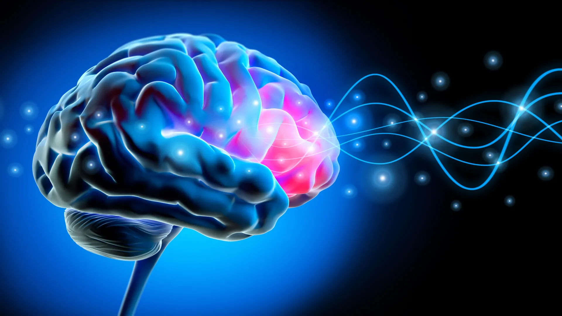
Deep brain stimulation (DBS) can significantly reduce Parkinson’s disease movement symptoms, and new research is helping scientists understand why the treatment works. Researchers found that DBS benefits may depend largely on activating specific brain networks that communicate through relatively fast beta rhythms, typically between 20 and 35 hertz (Hz).
The findings come from an interdisciplinary team of neuroscientists and clinicians from Cologne University Hospital, Düsseldorf University Hospital, Harvard Medical School, and Charité Berlin. Published in the journal Brain, the study, “Deep brain stimulus-response networks operate in the high-beta band in Parkinson’s disease,” is the first to combine electrophysiology and brain imaging—two research methods that have traditionally been studied separately.
Identifying how and where deep brain stimulation works
“For the first time, we have been able to characterize the DBS response network in Parkinson’s disease in both space and time,” says study leader Andreas Horn, professor of computational neurology at the University of Cologne. “We found that Parkinson’s disease can be treated most effectively when stimulation targets a precisely defined brain network. This network operates synchronously within a specific frequency band, helping explain why some patients respond well to deep brain stimulation.”
Deep brain stimulation of the subthalamic nucleus is an established treatment for reducing motor symptoms in people with Parkinson’s disease. The therapy uses surgically implanted electrodes to deliver controlled electrical pulses to deep areas of the brain.
Previous studies have provided only part of the explanation for how DBS works. Brain imaging research has helped identify where stimulation appears to be most effective, while electrophysiological studies have measured the frequencies of the electrical signals involved. Until now, researchers had not been able to examine the location and timing of these signals at the same time.
Mapping brain networks involved in Parkinson’s disease
To study this relationship, the research team analyzed data from a large, multicenter group of 50 patients, representing 100 brain hemispheres. The scientists simultaneously recorded brain activity using implanted DBS electrodes and magnetoencephalography (MEG).
These recordings allowed the researchers to map functional connections between deep brain structures and regions near the brain’s surface.
The analysis showed that an important network linking the subthalamic nucleus with frontal brain regions communicates primarily through relatively fast beta frequencies, ranging from 20 to 35 Hz. The strength of this connection was also associated with the degree of motor symptom improvement patients experienced after DBS electrode implantation.
How brain rhythms may influence DBS treatment response
“These findings suggest that specific brain rhythms may serve as communication channels between the subthalamic nuclei and the cerebral cortex, helping mediate the therapeutic effects of deep brain stimulation,” explains the study’s first author, Dr. Berne Berners of University Hospital Düsseldorf. “By targeting areas connected to these identified brain networks, clinicians may be able to personalize DBS settings more precisely in the future, particularly for patients who do not receive optimal relief from treatment.”
The findings could provide a foundation for tailoring deep brain stimulation to each patient’s individual brain networks, especially when standard DBS settings do not deliver the desired level of symptom improvement.
The researchers now plan to investigate more directly how deep brain stimulation changes activity within these brain networks. Further studies are underway to explore the causal relationships between brain rhythms, network connectivity, and treatment response.
This research was primarily funded by the Professor Klaus Tiemann Foundation.
Source: www.sciencedaily.com