


Human Brain Tissue Grows Inside Mice and Integrates With Their Nervous Systems
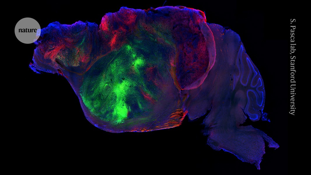
Human brain tissue transplanted into mice that lack a critical part of the brain can grow to fill most of the empty space and connect with the animals’ nervous systems, according to research results announced on September 16th.1
The study describes the most widespread integration of human brain cells into animals to date. The experimental approach could help researchers overcome a long-standing challenge in studying neuropsychiatric disorders using brain tissue grown in the laboratory.
Study co-author Serge Pasca, a neuroscientist at Stanford University in California, said the hybrid animals offer an unprecedented way to test drugs and investigate diseases involving abnormal brain development, such as cerebral palsy.
“We now have a very powerful new system” for understanding what makes the human brain unique and susceptible to disease, he says.
Creating human–mouse hybrids, or chimeras, can be uncomfortable for some people and raises ethical questions. Researchers in the field say that precautions are taken in studies such as this one. Pasca said the research underwent extensive oversight, including review by an independent bioethics committee.
Madeleine Lancaster, a developmental neurobiologist at the University of Cambridge in the United Kingdom who was not involved in the study, said the experiment was conducted a few days after the mouse pups were born, after the brain’s core wiring was already in place. This timing prevented the human cells from taking over complex thinking.
Behavioral tests also showed that the human tissue did not improve the rodents’ intelligence.
“The goal here is obviously not to make a super capable mouse, nor is it intended to be,” Lancaster says. The aim is to place human brain tissue in a realistic in vivo setting “so that we can begin to use it to understand human neurobiology and human neurological diseases.”
Why transplant human brain tissue into mice?
Neuroscientists have struggled for decades to study living human brain tissue. Human brain organoids—clusters of neurons grown from human stem cells in laboratory dishes—have opened a window into early neural development. However, organoids typically lack blood vessels and bodies that can send and receive signals.
In 2022, Pasca and his colleagues showed that when human brain organoids were transplanted into newborn rats, the neurons matured and connected to sensory pathways.2
Two years later, the researchers used those rats to assess the effectiveness of drugs called antisense oligonucleotides against Timothy syndrome, a serious genetic disorder associated with autism and epilepsy.3
In these early models, however, scientists placed the human grafts next to the rats’ existing, rapidly growing brain tissue. By the time the human neurons in the organoids began extending to establish connections, rodent brain cells had already occupied most of the available space.
Human cells fill an empty section of the mouse brain
In the latest study, Pasca and his team genetically engineered mice so that the precursor cells that form the cerebral cortex did not survive, creating an empty space in part of the brain.
“For me, real innovation is eliminating the race for space,” says Giorgia Cuadrato, a developmental neurobiologist at the University of Southern California in Los Angeles.
The human cerebral cortex is the wrinkled outermost layer of the brain involved in complex thinking and memory. Without competition from mouse cells, the transplanted human tissue expanded nearly fivefold within two to three months. It filled more than 90% of the empty space and sent protrusions deep into the rodents’ spinal cords.
Human tissue develops specialized neurons
The transplanted tissue developed into specialized neurons containing large, elongated cells similar to von Economo neurons. These cells, which are associated with social cognition in humans and some other animals, had not previously appeared in a laboratory dish.
“These cells are thought to be the cells most susceptible to neurodegenerative diseases, particularly frontotemporal dementia,” Pasca says.
Source: www.nature.com