


Why Are Marmoset Brains So Smooth? Study Reveals How Fewer Neurons Limit Brain Folding
Most primates have relatively large brains covered in distinctive folds and grooves. The common marmoset (Callithrix jacchus) is a rare exception: its brain is almost completely smooth, with very few folds. Scientists at the German Primate Center (DPZ), the Leibniz Primate Institute in Göttingen, have identified several cellular processes that help explain this difference.
Using miniature brain structures known as organoids, the researchers discovered that neural progenitor cells—the cells that produce neurons—develop differently in marmosets than in humans. These differences involve cell structure, the rate of cell division, and the timing of development. Together, they reduce the number of neurons produced, contributing to the marmoset’s smaller brain and smoother cerebral cortex. Published in Scientific Progress, the discovery provides new insights into primate brain evolution and may ultimately improve understanding of human brain development disorders.
Why Are Some Primate Brains Folded While Others Are Smooth?
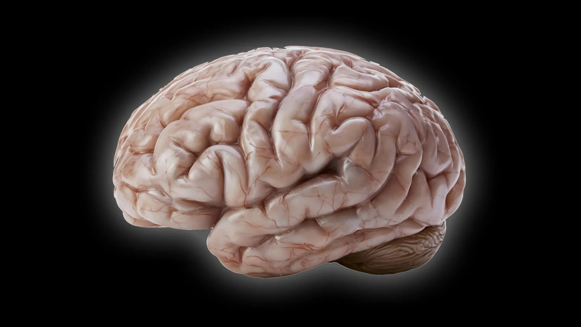
Brain size and the degree of surface folding are important characteristics that distinguish primate species. The human brain, for example, has a highly folded surface resembling the ridges and grooves of a walnut. This arrangement greatly expands the brain’s surface area, creating space for billions of neurons that support complex thinking and other advanced mental abilities.
Scientists believe the common ancestor of modern primates already had a moderately sized brain with at least some degree of folding. However, not all primates alive today retain these characteristics.
The common marmoset is a small monkey native to South America that is becoming increasingly important in biomedical research. Its brain has few visible folds, prompting researchers at the German Primate Center to investigate why it develops so differently from the brains of humans and other primates.
“We wanted to understand what changes at the cellular level inhibit marmoset brain growth and reduce fold formation,” explains Lidia Tinianskaia, one of the study’s two lead authors and a PhD student in the Junior Research Group Brain Development and Evolution at DPZ. “Early in development, the common marmoset brain exhibits the typical structure and organization of a largely folded primate brain. Therefore, as development progresses, processes that effectively slow down the production of neurons should occur.”
Scientists Identify What Slows Marmoset Brain Growth
To investigate these developmental differences, the research team compared brain organoids grown from human and common marmoset cells. The researchers focused on neural progenitor cells, which produce neurons during brain development.
These progenitor cells help determine the brain’s final size and shape. Producing more neurons can allow the cerebral cortex to expand and develop folds.
The researchers found that the smaller, smoother marmoset cerebral cortex is not the result of a single developmental change. Instead, several differences involving distinct types of progenitor cells work together to limit neuron production and brain folding.
“Our study showed that certain progenitor cells in the common marmoset divide significantly more slowly than in humans,” explains César Mateo Bastidas Betancourt, first author of the study and a PhD student in the Junior Research Group. “Other progenitor cells have a simpler structure and fewer processes than human progenitors, making them less proliferative. Both of these factors ultimately reduce the number of neurons, which contributes to the smaller size and less folding of the marmoset cerebral cortex.”
Scientists also identified important differences in when specific progenitor cell characteristics and behaviors emerge during development. Compared with humans, marmosets have a shorter overall period during which these cells can rapidly proliferate.
Together, the slower rate of cell division, simpler cell structure, and shorter period of rapid growth help explain why marmoset brains produce fewer neurons and develop far fewer folds.
Brain Organoids Reveal Important Developmental Clues
Much of the research relied on brain organoids: small, three-dimensional structures grown from cells that reproduce key features of early brain development. These miniature models allowed scientists to examine developmental changes across multiple samples and compare the timing of important processes.
“This study combines the advantages of in vivo and in vitro methods,” says Michael Heide, head of the Brain Development and Evolution Junior Research Group. “Such sample sizes are not feasible in primates, so organoids are well-suited for obtaining statistically robust results. We then repeated several key experiments in fetal brain tissue and confirmed our results from organoids.”
One particularly important finding involved the relationship between organoid development and actual marmoset brain development. The researchers found that marmoset brain organoids grown for 50 days closely matched the developmental stage of a natural marmoset brain at 90 days.
Establishing this relationship was essential because brain organoids and actual brains do not necessarily develop at the same rate. Identifying corresponding developmental stages allows scientists to make meaningful comparisons and interpret experimental results more reliably.
What the Smooth Marmoset Brain Reveals About Human Brain Evolution
The findings help explain how relatively small differences in the behavior and developmental timing of brain cells can contribute to major differences in brain size and surface folding among primates.
They also offer potential explanations for how different primate brain shapes and sizes emerged during evolution, including why some species developed relatively smooth brains while others evolved much larger, heavily folded brains.
Beyond their evolutionary significance, the findings provide a foundation for investigating how the human brain develops and what happens when those processes are disrupted. A deeper understanding of the cellular mechanisms that control neuron production and brain growth may ultimately help researchers study developmental disorders affecting the human brain.
Source: www.sciencedaily.com