


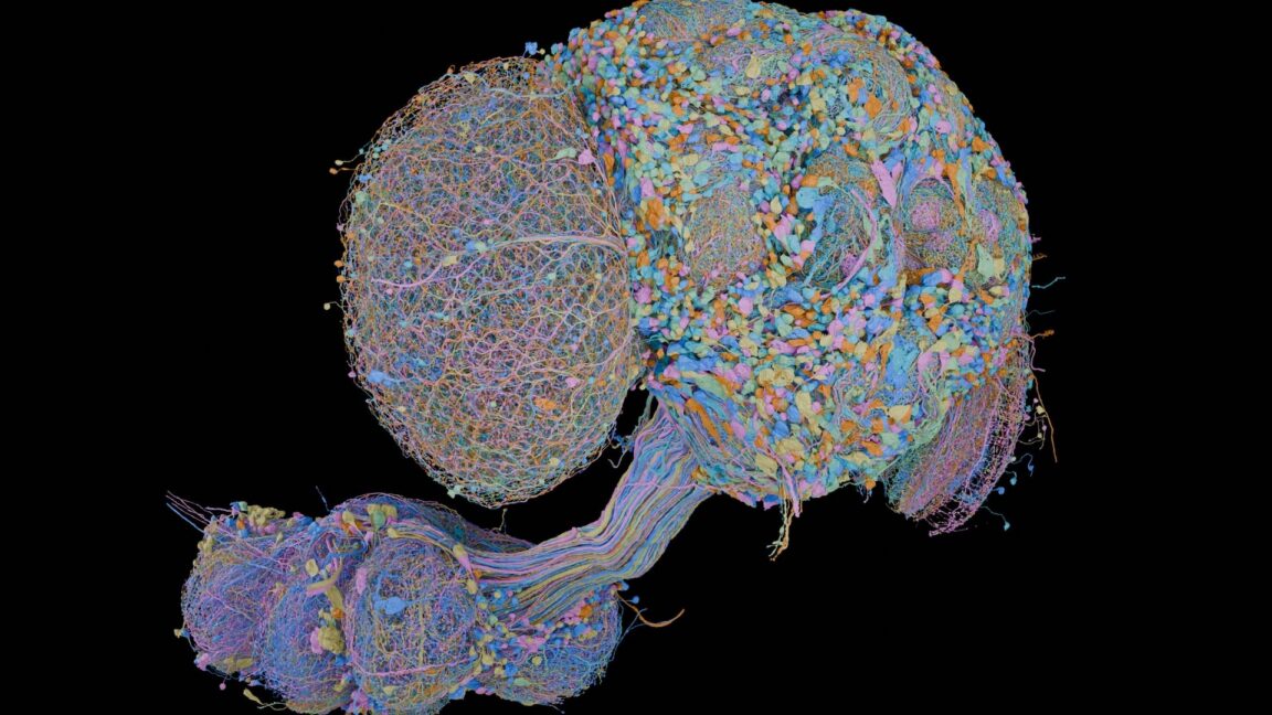
Every function of the brain depends on how neurons connect with one another. For instance, the visual system performs basic image recognition before sending information to the brain’s visual processing centers. When those centers identify text, they communicate with language-processing regions to interpret its meaning.
To understand how the brain processes information, scientists need a detailed map of its internal wiring. This map shows how information travels through different neural systems and is known as the connectome.
A connectome is a complete inventory of the neurons in a nervous system, including their three-dimensional locations and the connections they make with one another. These connections, known as synapses, make brain mapping a highly complex task. Individual neurons can extend multiple branching structures called axons and form hundreds of connections with other cells.
Although a fruit fly’s nervous system contains only about 150,000 neurons—and its brain includes just a portion of them—researchers identified more than 300 million synaptic connections in the fly brain. The result highlights the enormous challenge of creating a high-resolution connectome, even in relatively small organisms.
Mapping these connections has become easier as technology has advanced, but the process was once largely manual. Jerry Rubin, a senior group leader at the Janelia Research Campus and a senior author of the new study, described how researchers approached the problem decades ago while working at the UK’s Laboratory of Molecular Biology.
“When I arrived in 1971, they had already purchased a huge computer and developed the idea of using machine vision to build a nematode connectome,” Rubin said. The nematode is a small, transparent worm with just over 300 neurons, making it an apparently manageable model for studying neural connections.
However, the technology of the time was not powerful enough to complete the task. “It took me about two years to realize that computers weren’t powerful enough,” Rubin explained. “So I decided to print everything using photographic prints, rotate the neurons, and trace them by hand.”
Source: arstechnica.com