


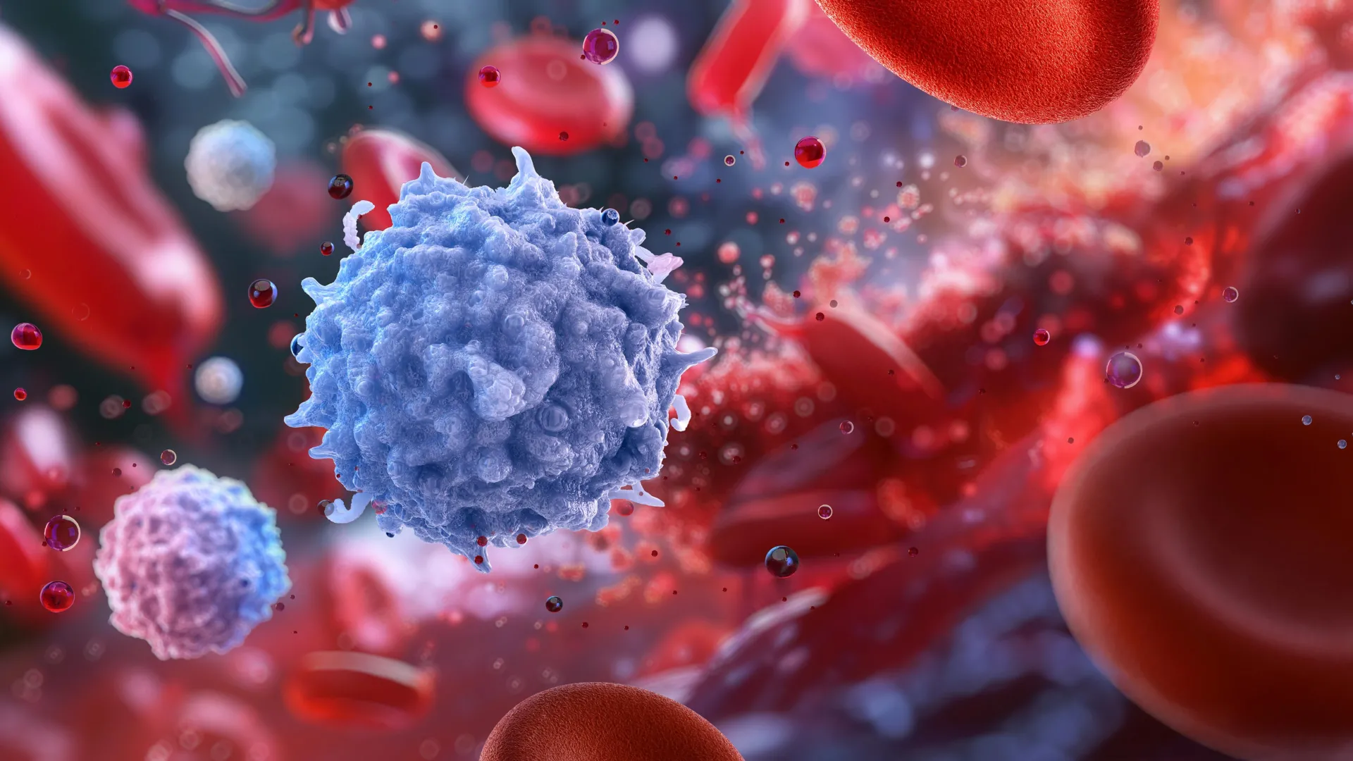
Researchers at Memorial Sloan Kettering Cancer Center (MSK) have solved a biological mystery that began more than 30 years ago, identifying the human gene DHRS7 as the source of an enzyme involved in the inflammatory signal 5-oxoETE. Experiments in zebrafish revealed that this pathway does more than attract immune cells to infections: it also protects healthy tissue from chemical damage caused by the immune response itself.
The findings, published October 7 in Nature, could inform research into cancer, inflammatory bowel disease (IBD), and asthma. They also suggest that treatments designed to strengthen the pathway might help tissues withstand inflammatory and oxidative stress, although further research is needed.
A mystery dating back to 1992
The investigation began in 1992, when researchers at McGill University studied 5-oxoETE, short for 5-oxo-6,8,11,14-eicosatetraenoic acid. This fatty signaling molecule helps direct immune cells toward sites of infection, acting as a chemical alert when the body needs to respond to a threat.
Scientists observed an enzyme that could both produce and break down 5-oxoETE. Although enzymes are proteins that facilitate chemical reactions, researchers could not identify the human gene responsible for producing this particular enzyme.
That gap made it difficult to manipulate the enzyme experimentally or determine how defects in its activity might contribute to disease. Researchers gave the enzyme a descriptive name based on its chemical function, but its genetic identity remained unknown for more than three decades.
The breakthrough came from Yanan Ma, PhD, a postdoctoral researcher at MSK’s Sloan Kettering Institute. Building on earlier work by former lab colleague King Lam Hui, PhD, Ma identified the human gene that produces the enzyme, allowing the team to investigate its function in detail.
When immune defenses damage healthy tissue
Ma works in the laboratory of cell biologist Philipp Niethammer, PhD. His team studies how cells respond to injuries, infections, and other forms of stress, as well as the roles of inflammation, tissue repair, and regeneration in cancer and other diseases.
When harmful bacteria enter the body, chemical signals recruit specialized white blood cells to the affected area. One of those signals is 5-oxoETE, which attracts immune cells capable of attacking invading microorganisms.
That response is essential for fighting infection, but it can also harm surrounding tissue. Immune cells release substances that destroy pathogens, and those substances can injure healthy cells if the response becomes excessive.
“They flood the site with toxic substances to kill the bacteria,” Dr. Niethammer says. “But this can cause damage to healthy cells if it gets out of control.”
Niethammer’s laboratory had spent years studying the molecular reactions involved in 5-oxoETE signaling. Researchers had begun to understand how the system’s components worked together, but the unidentified enzyme remained a critical missing piece.
Why zebrafish provided the crucial model
Mice and rats are widely used in biomedical research because their biological systems share many similarities with those of humans. However, they were not suitable for studying the complete 5-oxoETE pathway.
“Mice and rats are the primary model used in biomedical research,” says Dr. Ma, who received a Marie-Josée Kravis WISE Fellowship in 2023 for her work on the pathway. “It makes sense — they’re mammals, and there’s about an 80% overlap between their genes and ours.”
Despite that similarity, mice lack a known immune-cell receptor capable of recognizing 5-oxoETE. Without the receptor, researchers cannot examine the full signaling process in mice.
Zebrafish, by contrast, have retained the necessary biological machinery.
“This means that you can’t study the pathway in mice,” Dr. Ma says. “But the pathway is intact in fish. So picking the right alternative model was critical to advancing our understanding of how the human body responds.”
The zebrafish experiments enabled the researchers to study the signal in a living organism and eventually revealed a function that had gone unnoticed for decades.
Identifying DHRS7
Before turning to zebrafish, Ma and Hui searched for the enzyme’s genetic source in cultured human cells. They selected several candidate genes and systematically switched them off, looking for changes in enzyme activity.
One experiment produced a decisive result: when the researchers disabled DHRS7, the enzyme stopped functioning. The result established that DHRS7 was the gene scientists had been seeking since the early 1990s.
Additional tests using the rodent version of the gene showed almost no enzyme activity. That result was consistent with the absence of the 5-oxoETE receptor in mice and suggested that rodents had lost key functions associated with the signaling system.
Identifying DHRS7 solved the long-standing mystery and connected Ma’s work with a separate project in the same laboratory, led by postdoctoral researcher Miklos Lengyel, MD, PhD.
“Typically, when you have a paper with two first authors like Yanan and Miklos, there is just one story that two people contributed to equally,” Dr. Niethammer says. “But this paper is not like that. In this paper, there are two separate projects led by two different authors that came together as one larger discovery. And I think that’s extremely important to note.”
Removing the receptor made inflammation worse
Lengyel was using zebrafish to study colitis, an inflammatory condition affecting the colon. His experiments involved disabling selected genes to determine how they influenced intestinal inflammation.
In one experiment, he removed hcar1-4, the zebrafish version of the human gene OXER1. The gene produces the receptor that recognizes 5-oxoETE and enables cells to respond to its signal.
Because 5-oxoETE attracts immune cells and helps initiate inflammation, removing its receptor should theoretically have weakened the inflammatory response.
“So if you remove the receptor that detects the signal, you’d expect less inflammation, not more,” Dr. Lengyel says.
Instead, the zebrafish developed intestinal inflammation without an infection. The researchers initially considered whether neutrophils, the white blood cells normally recruited by the signal, were responsible. However, removing those cells did not eliminate the inflammation.
The result suggested that the 5-oxoETE pathway did more than activate immune defenses.
After Ma identified DHRS7, Lengyel disabled the gene in zebrafish, preventing production of the enzyme and disrupting the pathway. Based on 5-oxoETE’s established role in attracting immune cells, the researchers expected inflammation to decrease. Instead, inflammation increased, just as it had when the receptor was removed.
“It made absolutely no sense,” Dr. Lengyel says. “I had to prove using several different methods before the rest of the lab was convinced.”
The results indicated that the pathway also protects healthy cells. Disabling it removed not only an inflammatory signal but also an important mechanism that limits damage from the immune response.
An inflammatory signal that also acts as a shield
Further experiments showed how the protective function works. When immune cells attack microorganisms, they release highly reactive substances that can damage nearby healthy cells. The 5-oxoETE pathway helps tissues tolerate this chemical stress.
“This makes sense when you think about it,” Dr. Niethammer says. “These immune cells unleash chemical warfare against a pathogen, and organisms have evolved an elegant way of protecting healthy cells at the same time.”
Zebrafish intestinal cells became especially vulnerable when either 5-oxoETE or its Hcar1-4 receptor was absent. Even normal bacteria living in the intestine produced enough low-level chemical stress to cause problems when the protective system was disabled.
The researchers traced the protection to a group of enzymes known as NUDIX hydrolases. These enzymes help maintain cellular health by removing damaged DNA building blocks before they interfere with normal biological processes.
Without the 5-oxoETE pathway, intestinal cells could not withstand the chemical stress associated with ordinary gut bacteria. Some cells died, triggering inflammation even though no pathogen was present.
The findings show that the body uses the same molecular system both to recruit immune cells and to protect healthy tissue from the consequences of that response.
“As it turns out, this signal isn’t just an alarm, it’s also a shield,” Dr. Niethammer says.
Possible implications for cancer and inflammatory disease
The discovery could help explain how disruptions in 5-oxoETE signaling contribute to diseases involving excessive or poorly controlled inflammation. The pathway has been associated with cancer, IBD, and asthma.
Mutations affecting DHRS7 and OXER1, the human version of hcar1-4, have been identified in gastrointestinal and uterine tumors. Research has also linked mutations that prevent OXER1 from functioning properly with poorer outcomes in patients with colorectal, pancreatic, and kidney cancers.
The results raise the possibility that strengthening the pathway could help healthy tissues tolerate damaging conditions. One potential application would be protecting the intestine from oxidative stress associated with cancer treatment.
“More research is needed, but there’s a possibility that activating the pathway could increase the resilience of the gut to oxidative stress — which might help counteract damage caused by radiation therapy or chemotherapy,” Dr. Lengyel says.
The work may also be relevant to diseases affecting mucosal tissues, including the protective linings of the intestines and respiratory tract. In people with IBD, lower concentrations of 5-oxoETE in intestinal tissue have previously been associated with worse disease outcomes.
Asthma research points to another possible protective role. An independent study in primates found that blocking OXER1 weakened the immune-cell response but also caused protective mucus-producing cells in the airways to disappear.
Together, these observations suggest that efforts to suppress inflammation must account for the protective functions that some inflammatory signals perform. Whether this pathway can be targeted safely and effectively in humans remains uncertain.
Connecting infection, chemical stress, and physical injury
The researchers are also investigating how the pathway responds to physical damage, rather than only to infection or chemical stress. Doctoral student Zaza Gelashvili, MS, studies how cells detect mechanical forces associated with tissue injury.
When cells experience physical stress, changes in their membranes can initiate the same molecular chain reaction involved in inflammation. One early step occurs when stressed cell membranes release arachidonic acid, a fatty molecule used to produce 5-oxoETE.
Gelashvili, a co-author of the Nature study, led a separate investigation into molecular signals associated with physical wounds. That research was published in Nature Communications earlier in 2026. His work was also recognized with the 2026 Chairman’s Prize from the Gerstner Sloan Kettering Graduate School of Biomedical Sciences.
“There’s a mechanical component to the pathway that’s upstream of what Yanan and Miklos have been studying,” Gelashvili explains. “You can think of it as the first step in a chain reaction: Physical stress unlocks the raw material, oxidative stress drives the conversion, and DHRS7 amplifies the signal.”
The research suggests that the body combines information from different forms of cellular stress. Physical damage can initiate the process, chemical stress can influence the conversion of signaling molecules, and DHRS7 can strengthen the resulting signal.
“What we are really studying is a pathway that integrates two completely different kinds of stress — mechanical and metabolic — and converts them into a single, coordinated, protective response,” Dr. Niethammer says.
Study authors and funding
Additional researchers who contributed to the study were Yohannes Ambaw, Leehyeon Kim, Ritchie Ly, Siyang Peng, Meysoon Quraishi, Tobias Walther, and Robert Farese Jr., all affiliated with MSK.
Funding came from the National Institutes of Health (R35GM140883); an MSK Basic Research Innovation Award; a Starr Foundation Program for Discovery Science grant; a Tow Foundation fellowship; a Marie-Josée Kravis Women in Science Endeavor (WISE) fellowship; a Crohn’s Colitis Foundation Research Fellows Award (1268111); an Experimental Immuno-Oncology Scholars fellowship; and core facility services supported in part by MSK’s National Cancer Institute Cancer Center Support grant (P30CA008748).
Source: www.sciencedaily.com