


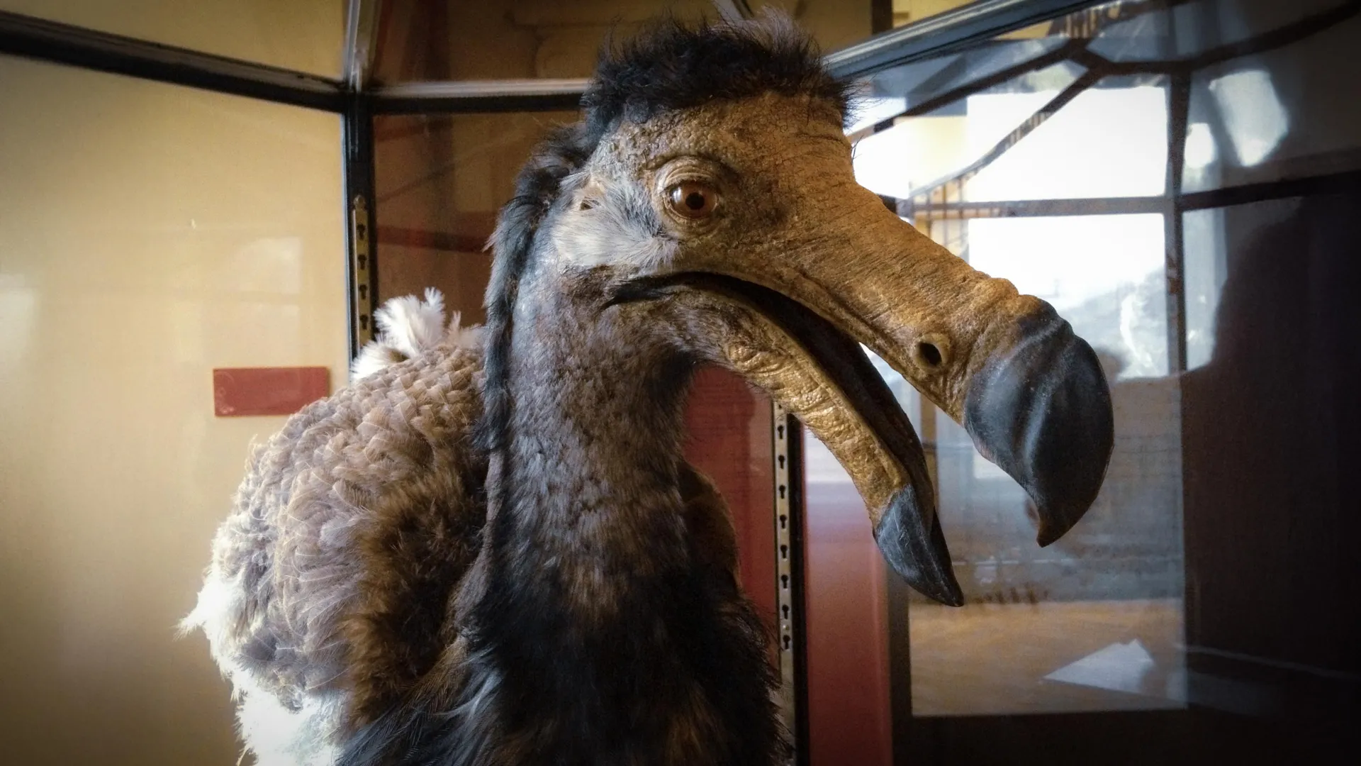
Only two complete dodo skulls are known to exist anywhere in the world. One is housed at the Natural History Museum Denmark, while the other is held by the Oxford University Museum of Natural History. The Copenhagen dodo skull has been part of the museum’s collection for centuries, and it has now helped scientists investigate one of the bird’s greatest mysteries: how the dodo sensed, explored, and interacted with its environment.
Using high-resolution CT scans and digital reconstructions of the space that once contained the dodo’s brain, researchers examined clues about the extinct bird’s sensory abilities. The findings suggest that the dodo experienced its surroundings differently from modern pigeons and doves, its closest living relatives.
The dodo may have followed a more flexible daily activity pattern, with movement and foraging potentially extending into dawn and dusk. It may also have possessed a stronger sense of smell and used its large upper beak to gather more tactile information from its surroundings. Together, these findings offer new insight into how the dodo may have searched for food and navigated the island of Mauritius before going extinct in the late 17th century.
“The dodo is one of the world’s most recognizable extinct animals, yet surprisingly little is known about how it actually lived,” says Peter Andrew Hosner, Associate Professor and Curator of Birds at the Natural History Museum Denmark and co-author of the study published in the Zoological Journal of the Linnean Society.
“Its behavior and foraging ecology have been debated for centuries. Having access to one of only two complete dodo skulls has given us a unique opportunity to compare the dodo with its closest living relatives and gain new insights into how it experienced and interacted with its environment,” he adds.
CT Scans Reveal the Dodo’s Hidden Brain Anatomy
The rare Copenhagen dodo skull has been displayed at the Natural History Museum Denmark for many years. Plans for a new museum building and permanent galleries, scheduled to open in 2027, created an opportunity to examine the specimen in unprecedented detail using high-resolution computed tomography, or CT, scanning.
The scans produced a highly detailed three-dimensional model without damaging the valuable fossil. Researchers used the model to reconstruct the skull’s internal structures, including the cavity where the dodo’s brain was located. As a brain develops, it can leave impressions on the surrounding bone. These preserved impressions, known as an endocast, can provide valuable evidence about how an extinct animal processed sensory information.
The study used CT data from three dodo skulls, including the only two complete specimens known to exist: one at the Natural History Museum Denmark and one at the Oxford University Museum of Natural History. Creating digital models of both complete skulls allowed the research team to compare them directly and identify anatomical features that were likely typical of the species.
“Studying extinct animals is a bit like detective work. Although the dodo’s brain disappeared centuries ago, it left an imprint on the inside of the skull. By digitally reconstructing those spaces and comparing them with the brains of living birds, we can begin to piece together how the dodo sensed, behaved, and interacted with the world around it,” says Christy Anna Hipsley, Associate Professor at the Natural History Museum Denmark and co-author of the study.
Evidence of Enhanced Smell, Touch, and Flexible Activity
Researchers compared the reconstructed dodo brain cavities with those of living pigeons and doves, which are the dodo’s closest surviving relatives. The comparison revealed a distinctive combination of sensory characteristics. The anatomy suggests that the dodo may not have been active exclusively during daylight hours and could have moved or foraged during the dim conditions of dawn and dusk.
The dodo may also have relied more heavily on its sense of smell than modern pigeons and doves. Its large upper beak appears to have been particularly well suited for detecting tactile information in the environment. Although anatomy alone cannot reveal the bird’s exact behavior, these findings provide important clues about how the dodo may have located food and explored its island habitat.
“The dodo’s skull is unlike that of any living bird, which makes it both fascinating and difficult to interpret. By comparing digital reconstructions of the world’s only two complete dodo skulls, we now have a much stronger foundation for identifying which features are truly characteristic of the species and what they can tell us about how the dodo lived,” Hipsley adds.
A More Complex Picture of the Dodo
Popular culture has often portrayed the dodo as clumsy or unintelligent, sometimes implying that its extinction was inevitable. However, the new findings do not suggest that the bird had reduced cognitive abilities compared with living pigeons and doves. Instead, the evidence points to a species with a unique combination of sensory adaptations that evolved over millions of years on the isolated island of Mauritius.
The researchers stress that skull anatomy cannot provide a complete picture of dodo behavior. Even so, combining CT data from multiple specimens—including the only two complete dodo skulls in existence—allows scientists to move beyond centuries of speculation and develop more evidence-based interpretations of the bird’s biology, senses, and ecology.
Peter Hosner says the study does not solve every mystery surrounding the dodo, but it provides a stronger framework for understanding how the extinct bird may have lived.
“The dodo has become a symbol of extinction, but in many ways it remains surprisingly mysterious. Every new piece of evidence helps us move beyond myths and build a more accurate picture of how this extraordinary bird lived before it disappeared,” he says.
Source: www.sciencedaily.com