


Algonquin Study Methods: Clinical Trial Design, γδ TCR Screening and Single-Cell Sequencing
This methods section describes the Algonquin clinical trial of belantamab mafodotin, pomalidomide and dexamethasone (BPd) in relapsed or refractory multiple myeloma (MM), along with patient sampling, cell culture, γδ T-cell receptor (γδ TCR) screening, single-cell sequencing, CapTCR-seq and statistical analysis.
Clinical Trial Design, Patients and Conduct
The Algonquin study is an ongoing, multicentre, open-label, single-arm, two-part study (ClinicalTrials.gov identifier NCT03715478) of BPd in patients with relapsed or refractory MM. Part 1 consisted of a dose-exploration phase22. Part 2 is evaluating the safety, tolerability and clinical activity of the dose and schedule identified in Part 1. Patients included in this interim analysis were enrolled in cohorts 2a and 2b, with the last patient enrolled on 14 September 2022. The clinical data cut-off date was 28 October 2025.
At screening, eligible patients with relapsed or refractory MM were aged ≥18 years, had an Eastern Cooperative Oncology Group performance status of 0–2, had undergone autologous stem cell transplantation or were considered transplant-ineligible, and had experienced disease progression after ≥1 previous lines of antimyeloma treatment. Patients were required to be lenalidomide-refractory and exposed to a proteasome inhibitor, either in separate regimens or in combination, and to have adequate bone marrow (BM), renal and cardiac function.
Patients with previous pomalidomide or BCMA-targeted therapy exposure, concurrent corneal epithelial disease other than mild punctate keratopathy, serious or unstable pre-existing medical conditions, psychiatric disorders, or other conditions—including laboratory abnormalities—that could interfere with safety, informed consent or compliance with study procedures were excluded. Sex was not considered in the study design or patient selection and was determined through self-reporting.
The Algonquin study was conducted at nine Canadian sites in accordance with the Declaration of Helsinki, International Council for Harmonisation Good Clinical Practice guidelines, applicable local regulations and institutional guidelines. All patients provided written informed consent. The study protocol, amendments and informed consent documents were approved by the University Health Network Institutional Review Board (REB numbers 16-5260, 18-5911 and 17-5357). Eligibility criteria, trial procedures, end points and participating sites have been described previously22.
Belantamab mafodotin (belamaf) was administered as a 30-min intravenous infusion using the following dose schedules: 1.92 mg kg−1 every 4 weeks in cohort 1; 2.5 mg kg−1 every 4, 8 or 12 weeks in cohorts 1a, 3a and 3b; total doses of 2.5 or 3.4 mg kg−1 every 4 weeks, split evenly with 50% administered on days 1 and 8 of each cycle, in cohorts 1b and 2, respectively; or a loading dose of 2.5 mg kg−1 on cycle 1 day 1 followed by 1.92 mg kg−1 every 4 weeks from cycle 2 onwards in cohort 1c. Pomalidomide was administered at 4 mg on day 21 of a 28-day cycle, and dexamethasone was administered weekly at 40 mg, or 20 mg in patients aged >75 years.
Patients remained on treatment until disease progression, unacceptable toxicity or withdrawal of consent. Ophthalmological examinations were performed before each belamaf dose, and preservative-free lubricant eye drops were required throughout treatment. Dose modifications were made independently for each drug according to predefined criteria based on the nature and grade of the adverse event.
Acquisition of Patient Samples
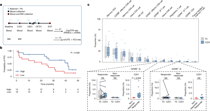
For the Algonquin patient cohort, BM and peripheral blood samples were collected at the time points indicated in Fig. 1a. Samples used in this investigation were selected and sequenced on an ongoing basis according to availability. BM samples were included for patients with matched baseline and cycle 2 day 1 samples, except for patient ALQ-19-019, whose baseline BM sample could not be sequenced because of poor viability. Peripheral blood samples were excluded only when poor sample quality prevented sequencing or for specific analyses, as indicated in the relevant figure legends.
For the distinct patient cohorts used to train PreGame—the discovery cohort and validation cohorts 1 and 2 (n = 22)—BM samples were obtained from patients with MM receiving standard-of-care BM aspirates at the Princess Margaret Cancer Centre or were purchased from commercial sources (iSpecimen and Discovery Life Sciences). Samples included untreated patients and patients who had received at least one previous line of therapy.
BM mononuclear cells were isolated using Ficoll according to the manufacturer’s recommended protocol (Sigma) and cryopreserved in fetal calf serum (FCS) containing 10% DMSO. Melanoma and lung cancer samples were obtained from surgical resections of patients receiving standard-of-care therapy at the Princess Margaret Cancer Centre. Tumour samples were digested using GentleMACS (Miltenyi) and cryopreserved as single-cell suspensions. All patient BM biopsy samples were obtained from the pelvic bone. Additional information on the sequencing cohorts is provided in Supplementary Table 1. All samples were collected with informed consent under protocols approved by the appropriate institutional review boards.
Cell Lines and Culture Conditions
The MM cell lines RPMI-8226 and MM1R were purchased from the American Type Culture Collection (ATCC), ALMC1 was purchased from Sigma, and INA6, JJN3, EJM and AMO1 were purchased from DSMZ. All MM cell lines were cultured in complete IMDM consisting of IMDM (Gibco) supplemented with 10% FCS, 2 mM l-glutamine, 1% non-essential amino acids, 1% penicillin–streptomycin, 10 µg ml−1 gentamicin and 55 µM β-ME. Human IL-6 (BioLegend) was added to the media for ALMC1 cells at 1 ng ml−1 and INA6 cells at 10 ng ml−1.
To generate CD1D-overexpressing myeloma cell lines, the human full-length CD1D construct was introduced into MM cell lines using lentivirus, and CD1D+ cells were purified by fluorescence-activated cell sorting (FACS). Jurkat-76 cells were obtained from N.H. and cultured in the same complete IMDM as the myeloma cell lines.
The melanoma cell lines A-375, A2058, 624Mel, 526Mel, 888Mel and WM3670; the lung cancer cell lines A549, H1568, H1573, H1437, H1792, H1944 and H2030; and the glioblastoma cell lines LN-229 and A-172 were cultured in complete DMEM (Gibco) containing 1% GlutaMAX (Gibco) and supplemented as described above.
The small-cell lung carcinoma cell line H69PR, mesothelioma cell lines H2452 and MSTO-211H, breast cancer cell lines HCC1143 and HCC1937, pancreatic cancer cell line ASPC1, and colon cancer cell lines LoVo and Colo205 were cultured in complete RPMI-1640 (Gibco) supplemented as described for complete IMDM. The pancreatic cancer cell line CFPAC-1 was cultured in complete IMDM.
Merkel cell carcinoma cell lines MCC14-2, MCC26, MS-1 and MCC13 were purchased from Sigma and cultured in RPMI-1640 supplemented with 20% FCS, 2 mM l-glutamine, 1% penicillin–streptomycin, 25 mM HEPES and 10 µg ml−1 gentamicin. Lenti-X 293T cells were purchased from Takara and cultured in complete DMEM as described above. Healthy primary cells used for screening were obtained from healthy donors or purchased from ATCC and Sigma and cultured according to the manufacturers’ recommended media and protocols. Tumour cell lines were confirmed to be free of mycoplasma.
Antibodies and Flow Cytometry
The following antibodies were used for flow cytometry: CD3 (OKT3; BioLegend, 317308 or 317322, 1:400); B2M (A17082A; BioLegend, 395716, 1:800); CD69 (FN50; BioLegend, 310910, 1:400); CD8 (RPA-T8; BioLegend, 301049, 1:400); CD4 (OKT4; BioLegend, 317420, 1:200); NGFR (ME20.4; BioLegend, 345132, 1:200); γδTCR (B1; BioLegend, 331210, 1:100); αβ TCR (IP26; BioLegend, 306720, 1:100); CD14 (61D3; Thermo Fisher, 53-0149-42, 1:50); CD2 (RPA-2.10; BioLegend, 300230, 1:75); CD235 A/B (HIR2; BioLegend, 306614, 1:200); CD138 (MI15; BioLegend, 356504, 1:100); CD319 (235614; BD, 750838, 1:20); HLA-ABC (W6/32; BioLegend, 311405, 1:50); HLA-A (1082C5; BD, 568024, 1:100); HLA-C (DT-9; BD, 566372, 1:100); BTN3 (232-5; BD, 566006, 1:100); Bw4 (REA274; Miltenyi, 130-123-760, 1:100); KIR3DL1 (DX9; BioLegend, 312707, 1:100); CD94 (HP-3D9; Invitrogen, 17-5094-4210, 1:100); GPR56 (CG4; BioLegend, 358205, 1:100); 4-1BB (4B4; BioLegend, 309809, 1:200); CD1D (51.1; BioLegend, 350305, 1:100); and CD1C (L161; BioLegend, 331505, 1:100).
Trustain Fc Block (BioLegend, 422302) was used to block Fc receptors. Fixable viability dye (Invitrogen, 65-0865-14), with or without Annexin V staining (BioLegend, 640920), was used to identify live and apoptotic cells. Unless cells were used for sorting or Annexin V staining, they were fixed with 4% paraformaldehyde before analysis. Flow cytometry data were acquired using a BD LSR Fortessa X20 or Beckman Coulter CytoFLEX instrument and analysed with FlowJo software (version 10, BD Biosciences).
Generation of B2M-Deficient NUR77 T2A–eGFP Knock-In Jurkat-76 Cells
Human NUR77 T2A–eGFP knock-in Jurkat cells were generated by modifying Jurkat-76 cells, an αβ TCR-negative derivative of the CD8− human T-cell lymphoma Jurkat cell line52. A CRISPR design tool was used to identify a unique single-guide RNA (sgRNA) targeting 5′-TTCCCAGGCAGGGGTCAGAA-3′ near the 3′ stop codon of the human NUR77 coding region. The sgRNA was chemically synthesized by Synthego.
A 1,761-bp homology-directed repair (HDR) template was designed with a 5′ homology region containing exon 8 of NUR77, a furin cleavage site (RRKR), a flexible linker (SGSG), a T2A (Thosea asigna virus 2A) ribosome-skipping peptide, an eGFP open reading frame and a 3′ homology region containing the NUR77 3′ untranslated region. To prevent CRISPR targeting of the HDR template, 17 of the 20 nucleotides in the NUR77 sgRNA target sequence were removed from the HDR sequence.
The double-stranded HDR DNA fragment was synthesized by Twist Biosciences. Sense and antisense single-stranded DNA HDR templates were prepared using a Guide-it Long ssDNA Production System version 2 kit (Takara Biosciences) and PrimeStar Max DNA polymerase with the following 5′ phosphorylated and unphosphorylated sense and antisense primers: NUR77_U1, 5′-CGTTTGTGACCCTGGGCAAGTCATTAGC-3′; and NUR77_L1, 5′-CTGTCATTTGTCTTCTTCCTAGAGTGAC-3′.
Ribonucleoprotein complexes were generated by mixing sgRNA with SpCas9 2NLS nuclease protein (both from Synthego) in electroporation buffer for 10 min at room temperature. Sense or antisense NUR77-T2A–eGFP HDR ssDNA template (1.5 µg) was added before electroporation of Jurkat-76 cells using an Amaxa 4D Nucleofector instrument (Lonza) with the CL-120 program. FACS sorting of eGFP+ cells enriched for successful HDR targeting, measured by NUR77 and eGFP expression after treatment with low-dose PMA–ionomycin (Invitrogen).
After establishing the cell line, B2M was knocked out by electroporating Cas9 and a B2M sgRNA (Supplementary Table 2). Knockout was confirmed by flow cytometry.
TCR Constructs
To normalize TCR surface expression, endogenous γδ TCR signal peptide sequences were mapped using the Department of Health Technology SignalP 6.0 signal peptide prediction service and replaced with the fibroin light-chain (FIBL) signal peptide sequence from Dendrolimus spectabilis. RefSeq and nucleotide sequence alignments from the NCBI database were used to create consensus sequence templates for human TCR γ2, 3, 4, 5, 8 and 9 with either constant region 1 or 2, as well as TCR δ1, 2 and 3.
Sequences were codon-optimized for expression in human cells using the GenSmart codon optimization tool from GenScript. To normalize expression, only amino acid differences in the paired γδ TCR framework 3 and framework 4 regions, together with the unique hypervariable complementarity-determining region 3 (CDR3) sequences from single-cell sequencing data, were appended to each expression template. A silent SacII restriction site was introduced into the region encoding Gly15 and Thr16 of the TCRδ constant region to facilitate cloning.
Approximately 1,500-bp T2A-linked γδ TCRs containing FIBL signal sequences were synthesized by Twist Biosciences and amplified using Q5 high-fidelity DNA polymerase (New England Biolabs, M0491L). The sense primer was XhoI_FiBL_GD_U1, 5′-ATTAGCGCTTCTCGAGGCCACCATGATGAGACCTATTGTG-3′, and the antisense primer was KpnI_DeltaC_L1, 5′-AGGCATGCCACGTTGGTACCATTTTTCATCACGAACAC-3′. Bold bases indicate engineered restriction endonuclease sites.
PCR products were gel-purified and cloned into a pTrans-NGFR-Puro expression vector using an In-Fusion HD Cloning kit (Takara Biosciences). The vector is based on the pcDNA3.1 backbone and contains an EF1A promoter with an intronic enhancer driving expression of the human TCRγ chain, followed by a furin cleavage site, flexible linker, T2A sequence, TCRδ sequence and BGH polyadenylation signal. A separate cassette contains the mouse Pgk1 promoter driving the extracellular and transmembrane regions of human NGFR, followed by a furin cleavage signal, P2A sequence and puromycin-resistance gene.
The EF1A–TCRγδ and PGK–NGFR–P2A–puromycin cassettes were flanked by the left and right terminal inverted repeat sequences of the Sleeping Beauty transposon system, as previously described53. Plasmids were transformed into Stellar chemically competent Escherichia coli cells (Takara Biosciences) and purified using a NucleoSpin Plasmid Transfection-grade kit (Macherey-Nagel, 740490). Plasmids were electroporated into Jurkat-76 NUR77-T2A–eGFP cells together with 5′ ARCA-capped SB100X Sleeping Beauty transposase mRNA.
Lentiviral γδ TCR expression plasmids were generated using a lentiviral backbone modified from pKLV2 (Addgene plasmid 67974). Each human FIBL–TCRγ–furin–linker–T2A–FIBL–TCRδ cassette was subcloned in-frame into an amino-terminal human NGFR–furin–linker–P2A cassette under the control of the 232-bp core EF1A promoter. Purified lentiviral plasmids were used to transduce primary T cells.
Sleeping Beauty SB100X ARCA-Capped mRNA Production
The hyperactive SB100X Sleeping Beauty transposase has approximately 100-fold higher transposition efficiency than the original SB10 and has been shown to enable efficient genetic modification of human T cells while avoiding many biosafety issues and time delays associated with retroviral and lentiviral approaches54. SB100X mRNA was used to enable rapid and stable insertion of γδ TCRs into NUR77-T2A–eGFP Jurkat-76 cells. These cells are αβ TCR-negative and express eGFP following TCR activation through the NUR77 locus.
The SB100X coding sequence from Addgene plasmid 12790955 was codon-optimized for expression in human cells, synthesized by Twist Biosciences and subcloned into pxInVitro, an in vitro mRNA expression vector. The vector contains a T7 promoter, mouse α-globin 5′ untranslated region, codon-optimized SB100X sequence and mouse α-globin 3′ untranslated region.
The pxInVitro SB100X vector was linearized at the 3′ end of the α-globin 3′ untranslated region using XhoI. The linearized plasmid was electrophoresed on a 1% agarose gel, excised and purified using a NucleoSpin Gel and PCR Clean-up kit (Macherey-Nagel, 740609). The complete T7–SB100X expression cassette was amplified using Q5 high-fidelity DNA polymerase with pIVT Fwd, 5′-TTGGACCCTCGTACAGAAGCTAATACG-3′, and pIVT Rev T120, 5′-T120CTTCCTACTCAGGCTTTATTCAAAGACCA-3′.
The amplified double-stranded DNA-tailed poly(T) T7–SB100X cassette was gel-purified and eluted in RNase-free NE buffer at 70 °C. A MEGAscript T7 In Vitro RNA Production kit (Ambion, Life Technologies, AM1334) was used to generate capped polyA+ transposase mRNA. Pseudouridin-5′-triphosphate and 5′-methylcytidine-5′-triphosphate were used to reduce mRNA degradation through cellular immune pathways57. A 5′ Anti-Reverse Cap Analog (ARCA 3′-O-Me-7G(5′)ppp(5′)G) was used to improve mRNA stability and translation58.
The T7 MEGAscript reaction was performed for 4.5 h at 37 °C, dephosphorylated with Antarctic phosphatase for 45 min at 37 °C and purified using a MEGAclear kit. Capped transposase mRNA was quantified using a NanoDrop ND-1000 spectrophotometer and assessed using native non-denaturing 1.2% agarose TAE electrophoresis with a single-stranded RNA ladder. Aliquots of 400 ng ml−1 5′ ARCA-capped transposase mRNA were stored at −80 °C.
Jurkat Cell-Based γδ TCR Screening
γδ TCR plasmids (1 µg each) and 1 µl of 400 ng µl−1 transposase mRNA were combined with 1 × 10⁶ Jurkat-76 NUR77-T2A–eGFP cells in pre-warmed Lonza electroporation SE buffer. Cells were electroporated using an Amaxa/Lonza 4D Nucleofector System with the CK116 program. After 48 h, puromycin (Invivogen) was added to enrich for TCR-expressing cells.
To screen for tumour reactivity, TCR-expressing Jurkat cells were mixed at a 1:1 ratio with MM cell lines and incubated for 16–18 h at 37 °C before flow cytometric analysis. The CD1C-restricted JR.2 TCR59 served as a negative control because the MM cell lines do not express CD1C (Extended Data Fig. 9l).
A tumour-reactive γδ TCR was defined as producing a ≥15% increase in the absolute number of GFP+ Jurkat cells compared with Jurkat cells cultured without MM cells. Activation strength was calculated as the percentage of GFP+ cells in the CD3+ Jurkat fraction cultured with an MM cell line minus the percentage in unstimulated Jurkat cells. For experiments involving healthy cells, the GFP+CD69+ double-positive fraction was used to calculate activation strength.
For some experiments with cell lines or healthy cells that did not express CD1D, the CD1D-restricted TCR 9C2 was used as a negative control60. Increases of 13–15% were considered borderline and excluded from bioinformatic analyses involving tumour-reactive T-cell populations. Increases of 0–13% were considered background, and the corresponding TCRs were classified as non-reactive.
The 13% non-reactive threshold was established from the activation-strength distribution of the negative-control TCR in the initial discovery cohort. It corresponded to approximately three standard deviations above the mean and exceeded the maximum observed value for the negative-control TCR across 98 measurements.
For patient-matched MM cells, BM mononuclear cells or CD138+ magnetically sorted tumour cells (Miltenyi) were mixed at a 1:1 ratio with Jurkat NUR77–GFP cells expressing the indicated γδ TCR. GFP expression was assessed after overnight co-culture. In some experiments, Jurkat NUR77–GFP cells expressing KIR3DL1 and/or CD8a were used, as indicated in the figure legends, and treated with KIR3DL1 antibody (clone DX9, 10 µg ml−1; R&D Systems). HMBPP (Sigma) was added at 1 µM to stimulate the Vδ2Vγ9 TCR. Validated MM-reactive γδ TCR CDR3 sequences are listed in Supplementary Table 3.
Lentivirus Production
Lenti-X 293T cells were cultured in DMEM containing 10% FCS, 2 mM l-glutamine, 1% penicillin–streptomycin and 1% GlutaMAX. Cells were seeded to reach 80% confluence after overnight incubation. The medium was replaced with viral production medium containing the same DMEM formulation with 0.1% penicillin–streptomycin.
Transfection mixtures were prepared in Opti-MEM with pre-warmed TransIT-lenti transfection reagent, 2 µg µl−1 psPAX2, 2 µg µl−1 VSVG and 2 µg µl−1 lentiviral backbone plasmid containing the gene of interest. Supernatants were collected 48 h after transfection, filtered through a 0.45-µm PVDF syringe filter and concentrated using Lenti-X concentrator according to the manufacturer’s protocol. Pellets were resuspended in viral production medium and stored at −80 °C in single-use aliquots.
Expansion of Tumour-Reactive γδ T Cells from Patient BM
Cryopreserved BM samples from patients with MM were thawed and flow-sorted to isolate viable γδ T cells that were double-positive or double-negative for CD94 and GPR56. Cells were sorted into IMDM containing 10% human serum, l-glutamine, 1% penicillin–streptomycin, 0.1% gentamicin and fungizone, together with irradiated K562 feeder cells expressing membrane-bound IL-21, CD48 and 4-1BB ligand.
Cultures also contained IL-2 (1,000 IU ml−1), IL-15 (20 ng ml−1) and PHA-L (2.5 µg ml−1, eBioscience). Cells were expanded for 7–10 days and then co-cultured with MM cell-line targets, either RPMI-8226 alone or a mixture of RPMI-8226, AMO1 and MM1R cells. Where indicated, cultures included 10 µg ml−1 anti-γδ-TCR blocking antibody (clone B1; BioLegend) or an isotype-control antibody (clone MOPC-21; BioLegend).
Tumour-cell apoptosis was assessed after co-culture by gating on CD138+ tumour cells. Cells positive for Annexin V or the viability dye were considered apoptotic.
T-Cell Cytotoxicity Assay
Human peripheral blood mononuclear cells (PBMCs) from healthy donors were purchased from StemCell Technologies. Untouched primary pan-T cells were isolated using a negative-selection magnetic separation kit (Miltenyi Biotec) and cultured in T-cell medium consisting of RPMI-1640 with 10% human serum AB, 1% penicillin–streptomycin and 0.1% gentamicin.
The gene encoding the αβ TCR was knocked out using a P3 Primary Cell 4D Nucleofector kit (Lonza) with the EH-115 program, Cas9 and an sgRNA listed in Supplementary Table 2. Following electroporation, cells were resuspended in T-cell medium and co-cultured with irradiated mOKT3 cells61 at a 10:1 ratio. IL-15 (10 ng ml−1) and IL-2 (20 ng ml−1) were added 24 h after activation.
At 48 and 72 h after initial activation, γδ TCR lentiviruses were added and cells were spinfected for 2 h at 1,000g and 32 °C. NGFR-expressing T cells were isolated 7 days after transduction using CD271 magnetic beads (Miltenyi Biotec).
For cytotoxicity testing, transduced primary T cells were co-cultured with luciferase-expressing RPMI-8226 cells at effector-to-target ratios of 2:1 or 0.5:1 for 18–20 h at 37 °C. Luciferase activity was then measured using a Steady-Glo Luciferase assay (Promega). Cytotoxicity was calculated from the difference in luminescence between RPMI-8226 luciferase cells cultured with or without γδ TCR-transduced T cells. To account for donor-specific background activity, the highest effector-to-target ratio for each donor that produced <35% background killing with the CD1C negative-control TCR was used.
γδ TCR Ligand Identification
γδ TCR-expressing, NUR77–GFP-expressing Jurkat cells were cultured for 16–18 h with MM cell lines in which B2M, BTN3A, HLA-A, HLA-C or total HLA-ABC had been knocked out using CRISPR. sgRNAs are listed in Supplementary Table 2. Cell lines were flow-sorted to achieve high purity, and disruption efficiency was confirmed by flow cytometry (Extended Data Fig. 9m).
To assess γδ TCR65 reactivity to different HLA-C alleles, artificial antigen-presenting cells expressing individual HLA alleles were generated as previously described43 and cultured with γδ TCR65 Jurkat cells. HLA-C*03:04 mutants were synthesized as gene fragments by Twist Bioscience and subcloned into pLenti plasmids to generate lentiviruses. These lentiviruses were used to stably express the mutants in RPMI-8226 cells in which endogenous HLA-C had been knocked out using CRISPR.
Preparation of Cells for Single-Cell RNA, CITE, TCR and BCR Sequencing
For the Algonquin clinical trial cohort, cryopreserved patient BM samples were thawed and viable CD2+, CD3+, CD14+ and CD34+ cells were isolated by flow cytometry. Before sorting, cells were labelled with TotalSeq-C Human antibody cocktail 1.0 (BioLegend) and additional TotalSeq-C antibodies against BCMA (C0056), CD138 (C0831) and FcRL5 (C0829).
For the distinct MM cohorts used to train PreGame—the discovery cohort and validation cohorts 1 and 2 (n = 22)—cryopreserved BM samples were thawed and viable CD3+ T cells and CD138+ and CD319+ MM cells were isolated by flow cytometry as shown in Extended Data Fig. 4a. Cells were labelled with TotalSeq-C Human antibody cocktail 1.0, antibodies against CD138, BCMA and FcRL5, and TotalSeq-C Hashtag antibodies for sample multiplexing.
Melanoma and lung samples were processed similarly with TotalSeq-C Human antibody cocktail 1.0. The CD3+ fraction was enriched from melanoma samples, whereas the CD45+ fraction was enriched from lung samples. Sorted cells were immediately used for single-cell library preparation with a 10x Genomics Chromium 5′ kit version 2 at the Princess Margaret Genomics Centre. γδ TCR libraries were prepared using two previously described γδ TCR enrichment primer sets, producing two distinct libraries for sequencing62,63. Libraries were sequenced using an Illumina NovaSeq 6000 sequencer.
Alignment of Raw Sequencing Data
Single-cell gene expression, surface protein, BCR and TCR data were processed using the 10x CellRanger pipeline (version 7.0.1) with the GRCh38-2020-A, TotalSeq-C Human Universal Cocktail version 1 399905 plus C0831, C0056 and C0829 barcodes, and vdj-GRCh38-alts-ensembl-7.0.0 references, respectively.
For multiplexed samples, raw gene-expression and CITE FASTQ files were first demultiplexed using cellranger multi, with feature_types = Multiplexing Capture specified for CITE FASTQ files. Demultiplexed BAM files were converted to FASTQ files using cellranger bamtofastq, with --reads-per-fastq set to the total number of reads obtained in the initial demultiplexing alignment.
Demultiplexed gene-expression FASTQ files were aligned together with CITE, BCR and TCR FASTQ files using cellranger multi, with feature_types = Antibody Capture and force-cells set to the number of cells determined from the first alignment. Multiplexed melanoma samples were processed similarly using CellRanger version 8.0.1. Non-multiplexed Algonquin samples were aligned using cellranger multi with default parameters. NSCLC samples were aligned using CellRanger version 6.1.2 and the cellranger count function.
Alignment of Raw γδ TCR Sequencing Data
Because γδ TCR alignment is not officially supported by 10x CellRanger and two primer sets were used for library preparation, a custom alignment pipeline was developed from previously described approaches1,62,63 (Extended Data Fig. 4b).
A custom VDJ reference was generated from the vdj-GRCh38-alts-ensembl-7.0.0 reference used for αβ TCR alignment. The reference was duplicated, and the regions.fa file was modified to convert TRG and TRD gene names into TRA and TRB gene names using the following commands: sed -i 's/TRG/TRA/g' regions.fa and sed -i 's/TRD/TRB/g' regions.fa.
Raw FASTQ files from each primer set—Mimitou (M) and Gherardin (G)—were independently aligned to the custom reference using cellranger multi with feature_types = VDJ-T. The raw FASTQ files were then aligned together using the same method. This produced three sets of γδ TCR sequences per barcode: M only, G only and GM combined. Where applicable, samples remained multiplexed.
TRA and TRB gene names were converted back to TRG and TRD, respectively, and barcoded TRG and TRD sequences were extracted separately from the filtered contig annotations file. To retain complete sequence information for downstream cloning and expression in modified Jurkat-76 cells while preserving CellRanger clonotype assignments, a clonotype key and a full-sequence key were created for each sequence.
The clonotype key contained V, D and J genes and the nucleotide sequence of the CDR3 region. The full-sequence key contained the complete TCR nucleotide sequence, including framework regions 1–4, CDR1–3 and gene usage. Where unique clonotypes differed in their full sequences, full sequences were concatenated to generate lists of unique clonotypes for each chain. TRG and TRD clonotype lists were joined to the clonotypes.csv alignment output to reconstruct γδ TCR pairing.
For each barcode, clonotype lists from the three alignments were merged. When alignments disagreed, the following decision tree was applied: paired clonotypes were selected over orphan clonotypes; paired clonotypes containing additional γ or δ sequences were selected over non-dual paired clonotypes; among paired non-dual clonotypes, source alignments were prioritized in the following order: present in G, M and GM alignments; present in G and M but not GM; present in either G or M alone; and present only in the combined GM alignment. Finally, the clonotype with the highest frequency was selected.
This custom pipeline maximized recovery of γδ TCR sequences from true γδ T cells while limiting the introduction of artefactual γ and δ pairs. Raw γδ TCR FASTQ files from each primer set were also aligned using the standard, unsupported 10x CellRanger method with feature_types = VDJ-T-GD and the inner-enrichment-primers sequences CCACAATCTTCTTGGATGATCTGAGACT and GTCCCAGTCTTATGGAGATTTGTTTCAGC. The number of unique paired γδ TCR sequences available for cloning and in vitro screening for each patient with MM is reported in Supplementary Table 4.
Quality Control, Normalization and Single-Cell Clustering
Quality control was performed at the individual-sample level before sample integration and re-normalization. Single-cell analyses were conducted in R version 4.2.1 unless otherwise indicated. For multiplexed samples, assignment-confidence tables generated by cellranger multi were used to remove blank or unassigned droplets, multiplets and cells assigned to other multiplexed samples.
Cells were further excluded if they were outliers for low surface-protein library size, expressed fewer than 200 features or had mitochondrial gene expression above 10%. Genes expressed in fewer than three cells were excluded. High-quality gene-expression libraries were converted into Seurat objects (version 4.3.0)64 and normalized with SCTransform (version 0.3.5)65 using method = “glmGamPoi”, vst.flavor = “v2”, variable.features.n = 3000 and vars.to.regress = “percent.mt”.
Cells were clustered using Seurat’s shared-nearest-neighbour algorithm with Louvain modularity optimization. Principal-component analysis and UMAP were used for dimensionality reduction. CITE data were normalized using the CLR method across cells as implemented in Seurat and clustered using the same approach. To jointly analyse gene-expression and CITE data, a weighted-nearest-neighbour graph was constructed and used for the final UMAP embedding (wnnUMAP).
BCR, αβ TCR and γδ TCR sequences obtained from VDJ alignment were added to Seurat objects after filtering for high-quality and, where applicable, confidently demultiplexed cells. Initial cell-type annotations were assigned using immune-receptor sequences, gene expression and protein expression of canonical lymphoid, myeloid and malignant-cell markers. Remaining low-quality clusters were identified and excluded.
After sample-level quality control, high-quality cells from samples in each sequencing cohort were merged and re-normalized. To account for batch effects, Harmony (version 0.1.1)66 was applied by batch or sample using a modest theta value of 0. Harmony and CITE embeddings were used to construct a weighted-nearest-neighbour graph and cluster cells. Sample-level cell-type annotations were used to limit overcorrection and preserve relevant biological variation.
Identification of Cell Types
Cluster-specific differentially expressed genes and proteins were identified using Seurat’s FindAllMarkers function, and cell-type annotations were refined relative to the sample-level annotations (Extended Data Fig. 4e).
Malignant MM cells were identified by clonal expression of the dominant BCR sequence in each sample and expression of SDC1, which encodes CD138, and TNFRSF17, which encodes BCMA. Cells with polyclonal BCRs and expression of CD79A and CD19 were annotated as normal B cells. Expression of an αβ TCR and canonical T-cell markers was used to identify T-cell subsets.
To classify cells as γδ T cells, γδ TCR expression, cluster membership, TRDV gene expression and other canonical T-cell genes were considered. Because γTCR genes can be rearranged and expressed in αβ T cells, cells were classified as true γδ T cells only when they contained a paired γδ TCR sequence or an unpaired γ or δ TCR sequence without a paired αβ TCR sequence. Cells without a γδ TCR sequence but with TRDV expression and close clustering within γδ T-cell clusters were also annotated as γδ T cells.
This approach identified two transcriptional γδ T-cell clusters marked by GZMB and GZMK expression and closely clustered with their αβ T-cell counterparts. Although NK cells expressed the γδ TCR constant genes TRDC and TRGC1, they were distinguished from γδ T cells by their lack of TCR sequences. Sparse myeloid-cell populations were annotated using canonical markers.
TCR Repertoire Diversity and Similarity
Single-cell TCR repertoire diversity was assessed using richness or Shannon diversity, where indicated. Repertoire similarity was measured using the Morisita–Horn index.
Richness was calculated as the total number of unique CDR3 sequences detected in a sample repertoire. Shannon diversity was calculated as \(S=-{\sum }_{i=1}^{R}{p}_{i}\mathrm{ln}{(p}_{i})\), where pi is the abundance of the ith TCR sequence. The Morisita–Horn index was calculated using the vegdist function with method = “horn” in the vegan package (version 2.6-4)67.
PreGame Development
PreGame was developed using multimodal features from gene expression, surface-protein expression and cell metadata. Only cells with confirmed tumour-reactive (TR) or non-tumour-reactive (NTR) γδ TCRs were included. Cells expressing γδ TCRs with borderline activation strength of 13–15% were excluded to reduce potential false positives.
Five feature categories were selected: a 19-gene expression signature; a five-protein surface-expression signature; S-phase score; G2/M-phase score; and cell-cycle stage (Extended Data Fig. 6a,b). Gene and protein signatures were identified from differential expression between TR and NTR γδ T cells. Cell-cycle signatures were calculated using CellCycleScoring in Seurat, and cell-cycle stage was label-encoded as a numerical feature.
The initial model was trained on the discovery cohort using a 27-dimensional feature matrix containing 88 TR cells and 361 NTR cells. To address class imbalance, synthetic minority over-sampling (SMOTE)68 was applied only to training splits. Because the data included continuous and categorical features, the SMOTENC function from the imbalanced-learn package (version 0.11.0) was used.
Tenfold cross-validation was performed by using nine subsets for training and one for testing in each iteration. Preprocessing, normalization and feature selection were performed before training splits, whereas oversampling was applied only to training data. The procedure was repeated ten times so that each cell was used at least once for testing. Predicted probabilities were recorded to calculate performance metrics, and the entire training and evaluation procedure was repeated 100 times to reduce sampling bias.
Model performance was assessed using the area under the curve (AUC), which guided hyperparameter selection. Of the machine-learning models evaluated, random forest models performed best and were selected for downstream analysis.
Differential Expression Analysis of Tumour-Reactive γδ T Cells
After in vitro screening of γδ TCRs from the discovery cohort was completed (Fig. 2b), TR and NTR labels were mapped to the single-cell data. Cells expressing γδ TCRs with borderline activation strength of 13–15% were excluded. TR cells (n = 88) and NTR cells (n = 361) were compared using FindMarkers in Seurat to identify differentially expressed genes and surface proteins for use as PreGame features.
Applying PreGame to validation cohorts 1 and 2 and performing subsequent reactivity screening increased the number of TR and NTR γδ T cells available for differential expression analysis. PreGame identified 1,616 TR cells and 374 NTR cells (Fig. 4a). This imbalance resulted from several highly expanded clonotypes (Fig. 4c), whereas the numbers of unique TR and NTR clonotypes were more comparable: 48 and 73, respectively.
To limit the influence of highly expanded clonotypes, clonotypes were randomly downsampled to 30 cells, where applicable, corresponding to twice the median number of cells per screened clonotype. FindMarkers was then used to identify differentially expressed genes and proteins. Cells with a sequenced BCR in addition to a γδ TCR, which probably represented rare doublets, were excluded before downsampling.
After 1,000 iterations, log2 fold changes, P values, false-discovery-rate-adjusted P values and expression values were averaged. Results with an average FDR-adjusted P value <0.05 were considered significant (Fig. 4e).
Clonotypes containing more than two cells were classified as expanded, whereas clonotypes containing one or two cells were classified as low frequency. Expanded and low-frequency cells were compared separately for TR and non-reactive clonotypes using FindMarkers. Genes or surface proteins were considered differentially expressed when the cell-level comparison produced an FDR-adjusted P value <0.05.
TCR Target Enrichment and Sequencing for Hybrid-Capture TCR Sequencing
Genomic DNA (76–500 ng) from peripheral blood buffy coat, fractionated PBMCs or fractionated plasma was used for library construction. DNA was mechanically sheared with a Covaris LE220 to produce fragments averaging approximately 250 bp. Fragmented DNA was ligated to duplex unique molecular identifier (dUMI) adapters containing dual-indexed 12-nucleotide barcodes and T-tailed overhangs (Integrated DNA Technologies).
Libraries were PCR-amplified using KAPA HiFi master mix for 4–6 cycles and enriched for the TRA, TRB, TRG and TRD loci using a custom xGen Lockdown probe set (IDT). Final pools were sequenced on an Illumina NovaSeq 6000 platform using 2 × 150-bp paired-end chemistry. Demultiplexing was performed using bcl2fastq version 2.20 to generate paired FASTQ files for each sample.
Alignment of Raw CapTCR-seq Data
Raw CapTCR-seq data were aligned as previously described27. Before alignment, barcode extraction and header tagging were performed to isolate and validate duplex barcodes embedded in the adapter sequences. The 12-nucleotide barcodes and flanking spacer sequences were removed from the 5′ end of both reads and inserted into FASTQ headers in the format @<readid>|<r1_umi>.<r2_umi>/1 or /2.
Barcodes were validated against a whitelist to support downstream error correction. Reads with mismatched spacers or invalid barcode sequences were excluded. TCR sequence alignment and analysis were performed using MiXCR version 4.6.069,70. The align step used the rna-seq preset with parameters optimized for partial alignments: -OallowPartialAlignments = true and -OvParameters.geneFeatureToAlign = VGeneWithP.
Two rounds of partial assembly were performed using assemblePartial, followed by extension with extend and full clonotype assembly with assemble. Final alignments were subjected to quality control. Samples were excluded when outlier total or aligned read counts resulted in an alignment rate below 50%. No samples from Algonquin clinical trial patients were excluded by this quality-control step.
TCR Clonotype Extraction and Repertoire Diversity Calculation
Final clonotypes for the TRA, TRB, TRG and TRD loci were exported together using exportClones with filters that excluded non-productive clonotypes: --filter-out-of-frames, --filter-stops, --dont-split-files and --export-productive-clones-only.
Total read fraction, representing abundance, was calculated as the sum of read fractions for each unique CDR3 sequence within each TCR grouping. TCR diversity metrics were calculated separately for each TCR locus and sample source—plasma or PBMC—using postanalysis individual with the following parameters: -f, --default-downsampling count-read-auto, --default-weight-function read and --only-productive.
MM Cell-Line Bulk RNA Sequencing and Transcriptional Clustering
Bulk RNA-sequencing data from 66 MM cell lines were previously generated by and obtained from the Keats Laboratory (Keats Laboratory data repository). FPKM counts were obtained, and genes with fewer than ten counts across all cell lines were filtered out. The remaining genes were used to construct a UMAP embedding in R using the umap package (version 0.2.10.0), with n_neighbors = 6, min_dist = 0.5 and otherwise default parameters.
Patient Survival Analysis
Progression-free survival (PFS) in the Algonquin clinical trial cohort was analysed using the Kaplan–Meier method. Numbers of patients at risk at specified months are shown below the survival curves.
For PFS comparisons between two groups, patients were stratified into high and low groups using the median value unless otherwise indicated. Statistical significance was assessed using log-rank tests for comparisons involving patients with BM sequencing samples and Gehan–Breslow–Wilcoxon tests for comparisons involving the entire cohort when many patients remained on trial.
Survival analyses were conducted in R version 4.2.1 using the survival package version 3.4-0, PHInfiniteEstimates version 2.9.5 and survminer version 0.5.0. Kaplan–Meier curves were generated using the ggsurvplot function in survminer.
Statistical Analysis and Data Visualization
Statistical tests were performed using R version 4.2.1 or GraphPad Prism version 10.6.1 unless otherwise indicated. Tests were selected as described in the figure legends. Results were considered statistically significant at P < 0.05.
Packages used for data visualization included tidyverse version 2.0.0, pheatmap version 1.0.12, ggplot2 version 3.4.2, ggalluvial version 0.12.5 and/or packcircles version 0.3.7, or as otherwise indicated.
Reporting Summary
Additional information on the research design is available in the Nature Portfolio Reporting Summary linked to this article.
Source: www.nature.com