


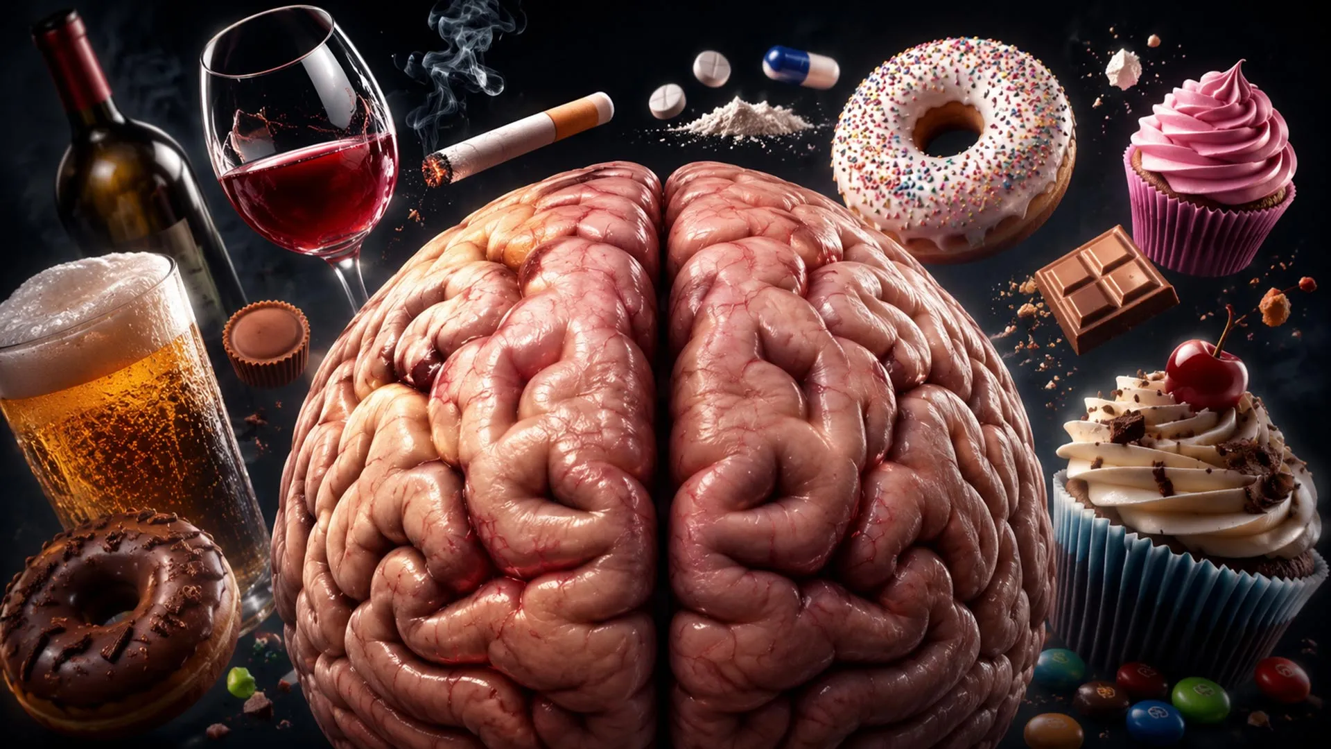
For many people, imagining a delicious burger or a cold beer creates a vivid mental image—and may inspire them to seek it out.
This connection between thought and behavior serves an important purpose: it motivates us to pursue the food, drinks, and other experiences we need or enjoy.
For some people, however, this process can become dysregulated. Repeated exposure to rewarding stimuli may contribute to substance-use and overeating disorders, including excessive alcohol consumption and food intake that can lead to obesity.
Research dating back to the 1970s has established a strong connection between mental imagery and drug-related behavior.
Understanding how cravings influence consumption is essential to understanding addiction. Although neuroscience has studied this relationship for decades, a new class of weight-loss medications may provide important clues about how the brain transforms thoughts about rewards into action.
These medications, including Ozempic and Wegovy, mimic the hormone GLP-1. They stimulate insulin release, slow digestion, and increase feelings of fullness. Known as GLP-1 receptor agonists, these drugs were originally developed to treat type 2 diabetes and help regulate blood sugar levels.
Weight loss became another notable effect. In clinical studies, people taking GLP-1 medications lost substantial amounts of weight—in some cases, amounts comparable to those associated with bariatric surgery.
However, GLP-1 drugs may have another, less widely recognized effect. Human studies suggest that GLP-1 receptor agonists can reduce alcohol intake. Preclinical animal research also indicates that these medications may reduce the use of several addictive substances, including cocaine, amphetamines, opioids, and nicotine.
These findings are changing how scientists understand the brain’s reward system. They may also lead to new treatment options for obesity, alcohol-use disorder, and dependence on other addictive substances.
How the Brain Regulates Rewarding Stimuli
Scientists have developed a strong understanding of the brain’s “reward circuitry,” a network that includes regions involved in producing and responding to the neurotransmitter dopamine.
Two key areas of this system—the ventral tegmental area (VTA) and the nucleus accumbens (NAc)—have been central to reward research for decades. Because these regions are closely associated with motivation and reinforcement, they were obvious candidates for investigating how GLP-1 drugs affect the brain. However, they contain relatively little GLP-1 receptor activity, making a direct mechanism less likely.
To understand how GLP-1 medications may reduce food and substance consumption, researchers must therefore examine other brain regions.
One potential candidate is the lateral septum, a structure located “upstream” from dopamine-producing regions. The lateral septum has traditionally been associated with emotional regulation and behavioral responses.
In 1953, American behavioral researchers Joseph Brady and Walle Nauta described “septal rage”. Animals with damage to the lateral septum displayed increased aggression, while direct stimulation of this region was associated with reduced aggression.
More recent research has placed the lateral septum at the center of a broader neural connectivity network, reshaping scientists’ understanding of its functions.
A connection between the lateral septum and the hypothalamus may help explain septal rage, but the lateral septum also interacts with other brain regions involved in a range of behaviors and functions.
The Brain’s Reward-Control Center
The lateral septum receives many of its primary inputs from the hippocampus.
The hippocampus is well known for its role in forming long-term episodic memories. Henry Molaison, widely known as Patient H.M., suffered severe memory impairment after surgery for epilepsy that damaged his hippocampus. He was unable to form new memories and appeared to live in a “perpetual present tense.”
The hippocampus also contains specialized neurons known as place cells. These cells fire in response to a person’s or animal’s location in space. Recent research suggests that the hippocampus also represents information about time.
This “where and when” information is transmitted to the lateral septum. Importantly, studies have shown that the lateral septum also contains place cells that respond strongly to rewards. In effect, these cells add information about “what is rewarding about this place” to the hippocampus’s representation of “when and where” an experience occurred.
The lateral septum then shares this information with dopamine-producing regions traditionally associated with the brain’s reward system.
Neuroscientists increasingly believe that the lateral septum helps the brain represent and evaluate rewards. It may allow us to mentally process rewarding experiences while communicating with the dopamine-based circuitry that generates motivation and feelings of satisfaction.
There is another reason to consider the lateral septum a potential target for the anti-consumption effects of GLP-1 receptor agonists: the region is rich in GLP-1 receptors.
Emerging research supports this possibility. One study found that activating GLP-1 receptors directly in the lateral septum reduced food consumption in mice. Another recent study reported a similar effect on alcohol intake.
My own laboratory also reported that GLP-1 drugs can reduce a specific type of activity in the lateral septum, potentially limiting its ability to communicate effectively with other brain regions.
These findings are reshaping our understanding of how the brain processes rewards and placing the lateral septum in the spotlight as a potential neural center for desire and craving.
Source: www.sciencedaily.com