


Scientists Discover the Human Brain May Have Two Ancient Developmental Origins
Scientists have long viewed the brain as a single organ with one common developmental origin. A new study led by Stanford Medicine challenges that assumption, suggesting that the brain is built from two distinct nervous systems that evolved separately over hundreds of millions of years.
For decades, the prevailing model held that the entire brain develops from one population of early progenitor cells. Under that view, the forebrain, midbrain, and hindbrain all trace back to the same developmental starting point.
The new findings suggest a different explanation: the human brain may be a combination of two ancient nervous systems. One system gives rise to structures responsible for essential functions such as breathing and heartbeat. The other develops into regions associated with language, mathematics, abstract thinking, consciousness, and reflection.
The discovery could also explain why scientists have struggled to grow certain brain cells in the laboratory. In particular, the findings may create new opportunities to study brainstem diseases such as spinal muscular atrophy (SMA) and amyotrophic lateral sclerosis (ALS), also known as Lou Gehrig’s disease.
“We show for the first time that the front of the brain develops from completely different progenitor cells than the back of the brain,” said Dr. Kyle Roe, associate professor of developmental biology. “Our findings mean we can grow neurons from the back of the brain, the hindbrain, in a Petri dish and study their function.”
The study was published in Nature Neuroscience on September 18. Loh is the senior author, while graduate students Carolyn Dundes and Rayyan Jokhai are co-first authors.
Two Distinct Origins of Brain Development
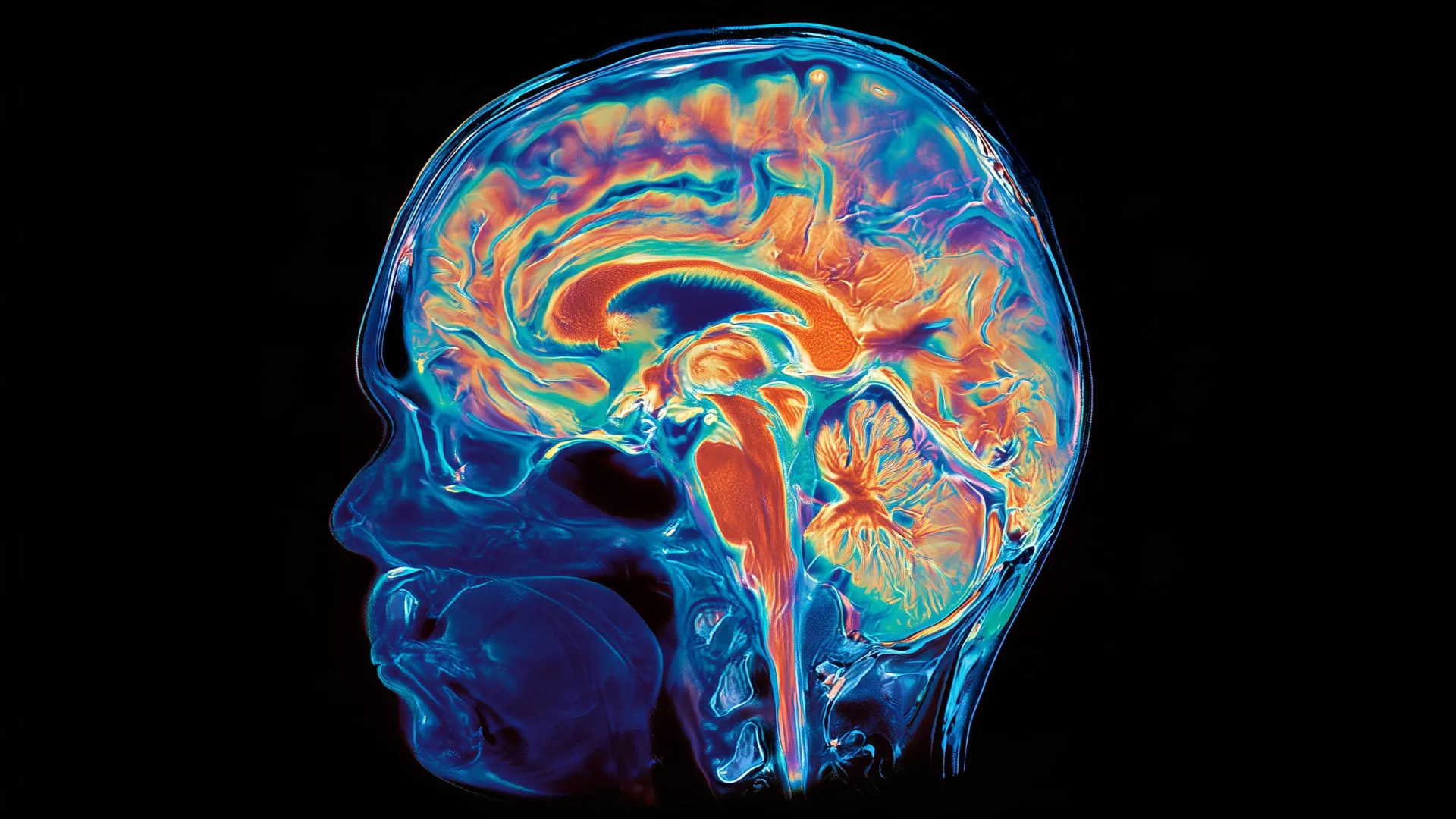
The adult brain is divided into three major regions: the forebrain, midbrain, and hindbrain. The forebrain supports many abilities associated with complex thought, including language, consciousness, and abstract reasoning.
The hindbrain is located toward the back of the skull and is often referred to as the brainstem. It controls automatic processes essential for survival, including breathing, sleep, heart-rate regulation, and hunger. Hindbrain neurons also send commands to the muscles of the face, tongue, and throat, which are necessary for speaking and swallowing.
Despite the hindbrain’s importance, scientists have struggled for decades to create human hindbrain neurons in the laboratory. This limitation has made it difficult to study diseases that affect the brainstem, including SMA and ALS.
SMA is the leading genetic cause of death in children under 1 year of age. ALS is often diagnosed between the ages of 40 and 70 and affects both the forebrain and hindbrain. In both conditions, certain hindbrain neurons gradually stop functioning. Patients may eventually lose the ability to swallow, increasing the risk of pneumonia if food or liquid enters the lungs, and may later lose the ability to breathe.
The Brain’s Developmental Divide Begins Early
The key insight came from examining one of the earliest stages of embryonic development. During gastrulation, the basic structures of the body begin to take shape.
Jokhai and Dundes found that the hindbrain does not emerge as a later branch of the developmental pathway that produces the forebrain and midbrain. Instead, it follows its own path from the beginning and develops in parallel with the other brain regions.
To investigate the process, the researchers studied developing mouse embryos and identified two distinct populations of brain progenitor cells.
One population expresses a gene called Otx2 and is destined to form the forebrain and midbrain. The second population expresses a gene called Gbx2 and develops into the hindbrain. Even at the earliest stages examined, the two populations remained separate and did not overlap.
The difference became even clearer when the researchers analyzed chromatin. Chromatin packages DNA inside cells and helps control which genes are accessible and which remain inactive.
The anterior neuroectoderm, which becomes the forebrain and midbrain, and the posterior neuroectoderm, which becomes the hindbrain, had fundamentally different chromatin compositions. These differences established separate developmental pathways from the outset.
“Previous attempts to create hindbrain neurons likely attempted to direct forebrain and midbrain progenitor cells into hindbrain cells, but our study shows that this is not possible,” Gjokaj said.
The finding may explain why earlier efforts to create hindbrain neurons were unsuccessful. Researchers may have been trying to convert one type of progenitor cell into another fate that it could not adopt.
“In stem cell biology, people are always obsessed with creating the final cell type, like neurons,” Gjokaj said. “But it’s important to start early in embryonic development. By paying careful attention to that early time point, we were able to find this fundamental divide in brain development.”
Researchers Grow Functional Human Hindbrain Neurons
Once the researchers understood that the hindbrain begins along a separate developmental pathway, they used that knowledge to address a major challenge in the laboratory.
For the first time, the research team succeeded in coaxing human pluripotent stem cells into functional hindbrain motor neurons. Pluripotent stem cells can produce every cell type in the human body.
The lab-grown cells behaved like real hindbrain neurons. They produced electrical signals known as action potentials and expressed proteins associated with hindbrain regions that control the muscles involved in facial movement and swallowing.
The researchers then examined evolutionary history to determine how far back the developmental divide might extend.
Evidence of Two Nervous Systems Dating Back More Than 500 Million Years
The researchers examined more than 550 million years of evolutionary history and found evidence of similar dual origins in chickens, zebrafish, and acorn worms. These small ocean-floor animals have a very distant common ancestor with humans.
The researchers also noted that jellyfish, which diverged from humans approximately 600 million to 700 million years ago, have two nervous systems located at opposite ends of their bodies.
Together, the findings raise the possibility that modern vertebrate brains developed when evolution brought two preexisting nervous systems into close physical proximity.
“Our study suggests that evolution acquired two pre-existing neural systems and pushed them spatially together,” Lo said. “It would probably be more efficient to have the brain as one organ, but we rely on this primitive method of making the brain as two separate parts.”
“Our findings were surprising because the word ‘brain’ probably suggests a continuous organ with a single origin,” Gjokaj said. “But even 500 million years ago, these separate nervous systems existed, and now they almost function as one, which is pretty amazing.”
Why the Discovery Matters for ALS and SMA Research
The research could have important implications for scientists studying SMA, ALS, and other diseases that damage the brainstem.
One major challenge is access to human tissue. Brainstem samples cannot be collected from living patients for routine research, making it difficult to examine the affected neurons directly. The ability to grow human hindbrain neurons in a dish could allow scientists to study these diseases with greater precision.
The hindbrain is also involved in weight management. It contains neural circuits that regulate hunger and is one of the systems affected by weight-loss drugs such as semaglutide.
Researchers now plan to investigate where the spinal cord originates during development and to better understand how SMA and ALS destroy hindbrain neurons.
“We now have a model to better understand these devastating diseases and work toward regenerative therapies for them,” Gjokaj said. “This is a very exciting new frontier in brain research.”
Researchers from the California Institute of Technology and the University of California, San Francisco, contributed to the study.
This research was supported by the National Institutes of Health through grants DP5OD024558, DP2GM146258, R00GM121852, R01DK115728, R01DE027538, T32GM119995, T32GM007365, T32GM007790, and F31DE031154; the National Science Foundation; the California Institute for Regenerative Medicine; the Spinal Muscular Atrophy Foundation; a grant from the Stanford Maternal and Child Health Research Institute; the Stanford Beckman Center and Ludwig Center; the Siebel Stem Cell Institute; the Steinhardt-Reid Foundation; the Gatsby Charitable Foundation; the Howard Hughes Medical Institute; the Packard Foundation; the Pew Charitable Trusts; the Baxter Foundation; the Human Frontier Science Program; and the anonymous Fickell, Gilbert, and Steinhardt-Reid families.
Source: www.sciencedaily.com