


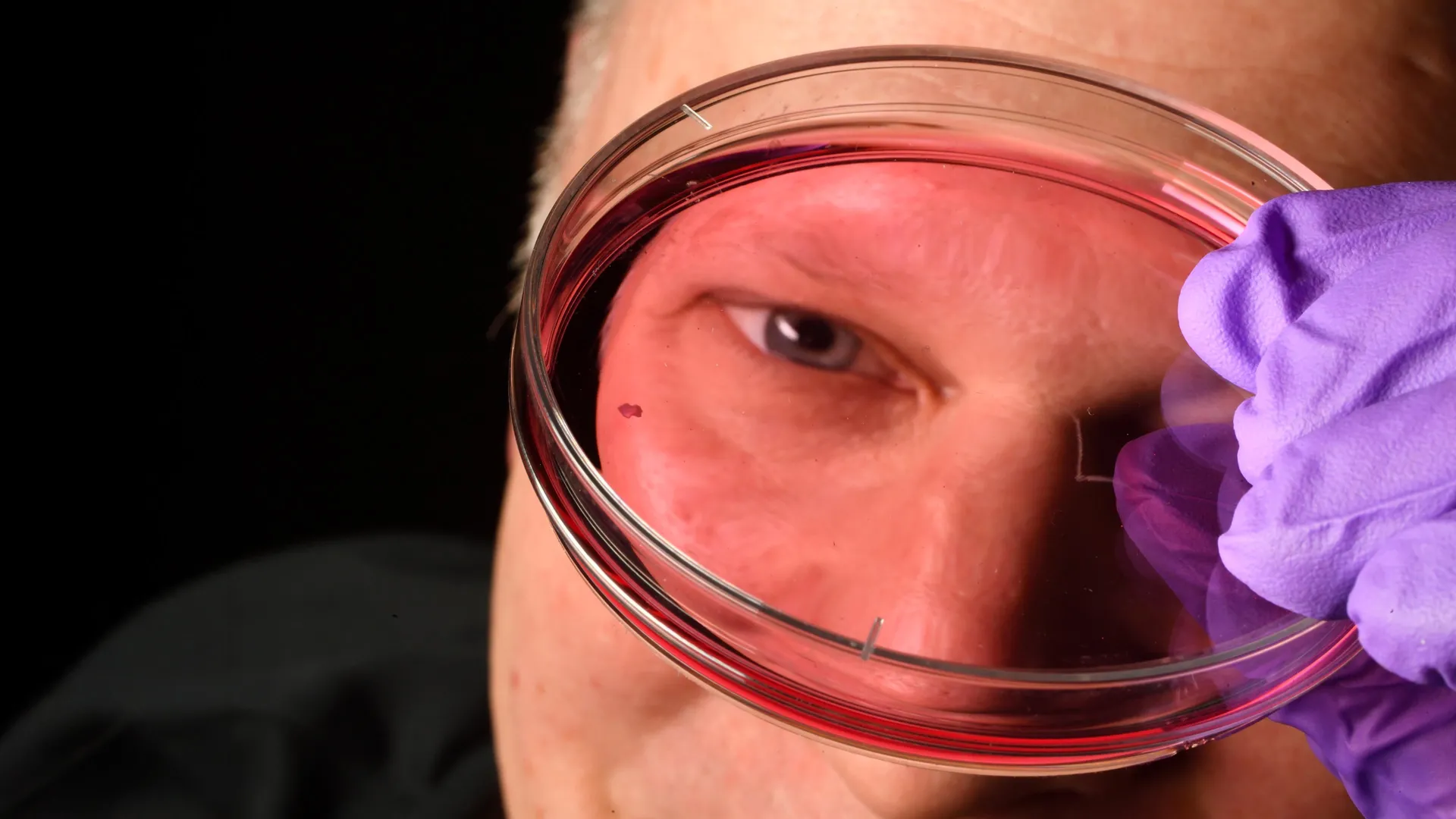
Researchers at Johns Hopkins University have revealed groundbreaking insights into the development of sharp central vision in humans before birth. Their study identified crucial interactions between vitamin A-derived molecules and thyroid hormones in the retina, challenging long-standing theories about the formation of light-sensing cells. This discovery has the potential to inform future treatments for conditions such as macular degeneration and glaucoma, which severely affect vision.
The study, published in Proceedings of the National Academy of Sciences, utilized lab-grown retinal tissue to explore these critical interactions.
Lab-Grown Retina Reveals Mechanisms of Clear Vision
“This is a major step toward understanding the central retina, a vital area of the eye that is often the first to deteriorate in individuals with macular degeneration,” said Robert J. Johnston Jr., associate professor of biology at Johns Hopkins University and leader of the study. “By advancing our understanding of this region and creating organoids that replicate its functions, we aspire to one day cultivate and transplant these tissues to restore vision.”
To study the development of the human eye, researchers employed organoids—small clusters of tissue cultivated from fetal cells that closely mimic retinal structures. Over several months, they monitored these lab-grown retinas to unravel the cellular events that shape the fovea, the region at the center of the retina responsible for sharpest vision.
This research emphasized the role of cone photoreceptors, the light-sensitive cells crucial for daytime and color vision. These cones can be classified as blue, green, or red, each type reacting to different wavelengths of light. Although the fovea comprises a small area of the retina, it accounts for roughly half of human visual perception. Notably, the fovea exclusively contains red and green cones, in contrast to the rest of the retina, which houses all three cone types.
Remarkable Transformations in Cone Cells
Humans possess a unique combination of three cone types, enabling a vast spectrum of color vision, yet how this specific arrangement develops has remained elusive for many decades. Previous attempts to study this phenomenon faced challenges, as common research animals like mice and fish do not exhibit the same photoreceptor configuration, according to Johnston.
This new discovery indicates that the foveal cone arrangement is established through a series of well-timed events during early fetal development. Blue cones begin to arise around 10 to 12 weeks of gestation, but by 14 weeks, they transition into red and green cones.
The researchers pinpointed two distinct mechanisms responsible for this transformation. Initially, retinoic acid—a molecule derived from vitamin A—is degraded, limiting the production of new blue cones. Subsequently, thyroid hormones facilitate the conversion of the remaining blue cones into red and green cones.
“Retinoic acid helps to establish the pattern, while thyroid hormones are crucial for transforming the remaining cells,” Johnston explained. “This transformation is significant, as the presence of blue cones can impair vision.”
Revising Established Theories
The findings offer a fresh perspective on a question that has baffled vision researchers for decades. Traditionally, it was believed that blue cones formed in the center of the retina and later migrated outward. In contrast, the new evidence suggests these cells remain stationary but change their identity into red and green cones, creating the specialized structure necessary for optimal vision.
“About 30 years ago, the prevailing model suggested that some blue cones in that area somehow relocated, influencing the future development of these cells while retaining their original identity,” Johnston remarked. “While we can’t completely dismiss this older model, our data strongly supports a different narrative: these cells actually transform over time, which is quite surprising.”
Potential for Future Vision Restoration
Researchers are optimistic that these findings may pave the way for novel strategies in treating vision loss. Johnston’s team is actively refining retinal organoids that more accurately replicate the functionalities of the human retina. Improved models could aid scientists in generating healthier photoreceptor cells for future cell-replacement therapies targeting diseases like macular degeneration, which currently lack effective treatments.
“The ultimate aim of employing this organoid technology is to develop nearly custom-tailored photoreceptors,” stated Hussey, now a molecular and cell biologist at CiRC Biosciences, a cell therapy firm based in Chicago. “These are extensive, long-term experiments requiring optimization for safety and efficacy before clinical application. Nevertheless, it represents a promising path forward.”
Source: www.sciencedaily.com