Scientists Explain How Ancient Human Brains Survive for Thousands of Years
The discovery of remarkably well-preserved human brains among otherwise skeletal remains has puzzled archaeologists and pathologists for decades. New research now suggests that ancient brain preservation is not a rare accident, but the result of a distinct chemical process triggered by decay.
“We found that brain preservation isn’t a rare anomaly — it’s a novel chemical pathway,” first study author Alexandra Seviour, a doctoral researcher in paleobiology at the University of Oxford, told Live Science in an email. The study, published June 19 in the Journal of Proteome Research, found that the same reactions that normally break down brain tissue can, under certain conditions, bind proteins together and make them highly resistant to further decay.
Although the brain is among the first organs to begin decomposing after death, it is surprisingly common in the archaeological record. More than 4,400 preserved brains have been identified in human remains dating back as far as 12,000 years, according to earlier research by Seviour.
Latest Videos FromLive Science
Mummification, freezing and saponification — a process in which body fat becomes a waxy substance known as grave wax — can preserve soft tissue for centuries or even millennia. These processes generally protect several parts of the body, including organs, skin and muscle. However, about one-third of the ancient brains found by archaeologists do not fit this pattern. In these cases, a shrunken mass of protein is the only soft tissue remaining among the bones.
Most of these unusual discoveries come from waterlogged, oxygen-poor environments, including riverbeds, lake shores, flooded caves and shipwrecks. “Water, being nature’s solvent, is typically associated with decomposition, not preservation,” Seviour said. “So the surprise is really the selectivity.” The central mystery is why the brain survives when other soft tissues disappear.
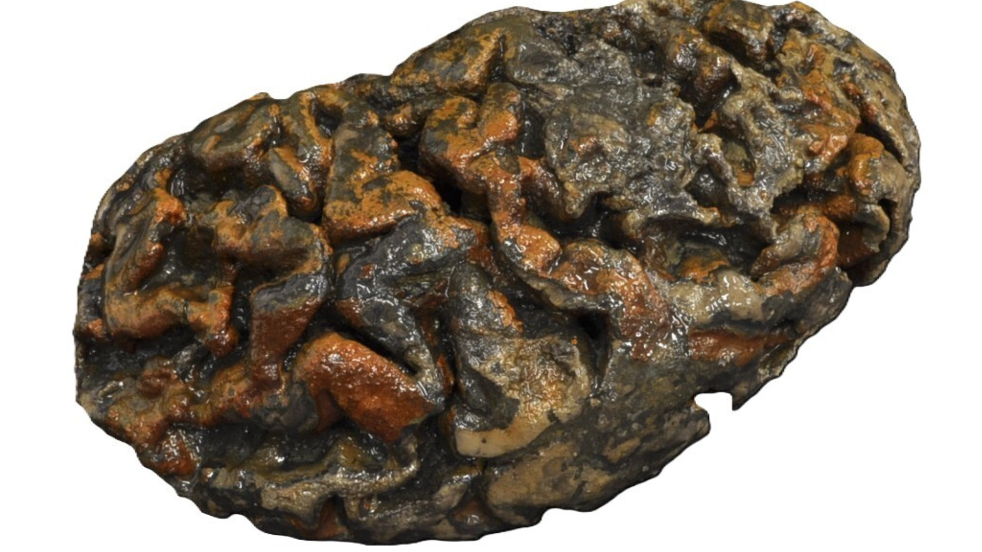
The preserved brain of an adult whose burial was found in Bristol. The brain is coated with clay from a waterlogged grave.
(Image credit: Alexandra Morton-Hayward)
How researchers tested ancient brain preservation
Seviour’s team proposed that waterlogged, oxygen-depleted burial sites interact with the brain’s distinctive structure and chemistry. Rather than allowing the tissue to break down completely, these conditions may redirect decomposition and stabilize the brain’s proteins through a different chemical pathway.
To test the idea, the researchers buried mouse carcasses under four combinations of water and oxygen levels. They then tracked how the brains decomposed over six months.
“At 24 hours, 72 hours, one week, six weeks, three months and six months, we dissected the brains and analyzed them using high-resolution mass spectrometry,” Seviour explained. The team examined which proteins remained, which disappeared and what chemical changes occurred in the surviving proteins.
Get the world’s most fascinating discoveries delivered straight to your inbox.
The analysis produced more than 1.26 million protein-decay trajectories. These results allowed the researchers to identify when decomposition changed under different environmental conditions. During the earliest stages, decay followed similar patterns in all samples. After several weeks, however, oxygen levels became the main factor.
High-oxygen conditions accelerated the breakdown of brain proteins. In contrast, wet and oxygen-poor conditions encouraged the formation of hardened protein structures that resisted further decomposition. This process helped preserve the remaining brain tissue.
Free radicals may hold the key
The researchers attribute this unusual preservation process to free radicals — highly reactive particles containing an unpaired electron. In oxygen-rich environments, free radicals can trigger a chain reaction that rapidly destroys the structure of brain proteins.
In oxygen-depleted environments, there is not enough oxygen for the same destructive cascade to continue. Instead, chemical intermediates can form crosslinks with nearby parts of brain proteins. These crosslinks produce tough, insoluble aggregates that are difficult for microbes and other decay processes to break apart.
Brain tissue may be especially suited to this self-limiting preservation pathway. It contains metals that promote free-radical reactions, membranes where reactive molecules can accumulate and “redox-active” amino acids capable of absorbing free radicals and forming crosslinks. The skull may also contribute by limiting the movement of fluids and oxygen around the brain.
Richard Evershed, an organic geochemist at the University of Bristol who was not involved in the study, praised the researchers’ detailed analysis. He said it would be valuable to investigate whether similar processes affect proteins in other archaeological tissues.
“Comparing more tissues — including other organs and muscles — would be really useful,” Evershed told Live Science. Such comparisons could show whether the preservation process is unique to the brain and help explain how proteins survive in other archaeological materials, such as pottery residues and dental calculus.
What ancient brains could reveal about neurodegenerative disease
The findings could have implications beyond archaeology. Seviour said the molecular signature of the decay-resistant peptides resembles patterns associated with neurodegenerative diseases such as Alzheimer’s disease.
Future research will examine how closely the two molecular signatures match. If the similarities are confirmed, preserved ancient brains could offer new insights into how neurodegenerative diseases develop and progress.
Morton-Hayward, A., Flannery, S., Berry, P., Vendrell, I., Johansen, A., Hansen, M., & Fischer, R. (2026). Molecular solution to the paradox of ancient brain preservation. Journal of Proteome Research.
Test your knowledge of the most complex organ in the human body with our brain quiz!