


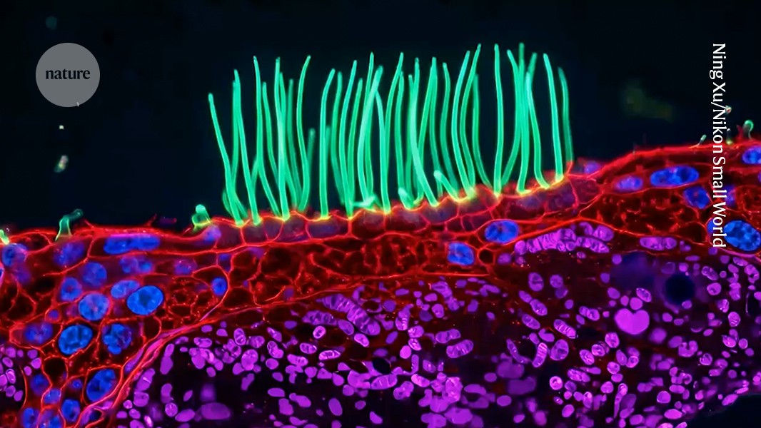
Credit: Ning Xu/Nikon Small World
AI Debate Erupts Over Award-Winning Microscopy Video
An award-winning microscopy video has sparked a debate over whether artificial intelligence can alter the scientific meaning of experimental images. Researchers say the winning entry in this year’s Small World in Motion competition appears to depict biologically impossible features that may have been introduced or hallucinated by AI-assisted processing.
“Scientific images are more than just illustrations,” says Melanie White, a developmental biologist at the University of Queensland in Brisbane, Australia. “They are data and we need to be able to trust that what we are seeing is based on the underlying measurements.”
Markus Sauer, who studies super-resolution microscopy at the University of Würzburg in Germany, says using AI tools to visualize experimental data is not automatically problematic. The concern arises when AI-generated images or videos misrepresent, exaggerate or alter experimental results, he says.
Microscopy competition winner used AI-assisted post-processing
On September 15, Ning Xu, an optical engineer at the National University of Singapore, was named the winner of an annual competition run by Nikon Instruments. The contest recognizes videos captured using optical microscopes.
Xu’s entry shows the movement of hair-like structures called cilia, which cover parts of the lungs. The tissue sample came from a child with primary ciliary dyskinesia, a rare genetic disorder that causes chronic lung, sinus and ear infections.
In the video, the cilia move over red, purple and blue structures. These structures were not described in the contest website’s original blog post about Xu and the video.
Researchers question the biological accuracy of the video
Days after the announcement, microscopists began questioning the accuracy of the clip on social media. Edward Phelps, a bioengineering researcher at the University of Florida in Gainesville, wrote on LinkedIn that the purple structures resembled mitochondria, but that subepithelial structures made up of extracellular mitochondria and measuring the same size as a cell nucleus do not exist in biology.
Phelps added that although the blue structures resemble atomic nuclei, they do not behave like them. He also said it was unclear what the red stain represented.
Nikon discloses use of an “unsupervised” AI model
On September 22, Nikon Instruments updated its blog post about Xu’s entry to state that he used an “unsupervised” AI model to assist with “post-processing.”
Xu did not respond to Nature’s request for comment about the researchers’ questions regarding the biological accuracy of the video. However, in response to concerns posted on LinkedIn, he said his team used post-processing to enhance the visual representation of the region beneath the cilia “without making any anatomical claims about what the rendered features represent.”
Xu said no AI was used to generate the cilia or their movements. Instead, the model was applied to reconstructed grayscale data to distinguish and colour structures with similar morphology.
The video was created for a competition celebrating the beauty of microscopy. “We wanted the final presentation to be not only scientifically interesting, but also visually appealing,” Xu wrote.
How faithfully does the video represent the microscope data?
White says it remains unclear how faithfully the video represents the information captured by the microscope. She says AI tools may have introduced biological structures into the data that were not actually present.
Source: www.nature.com