


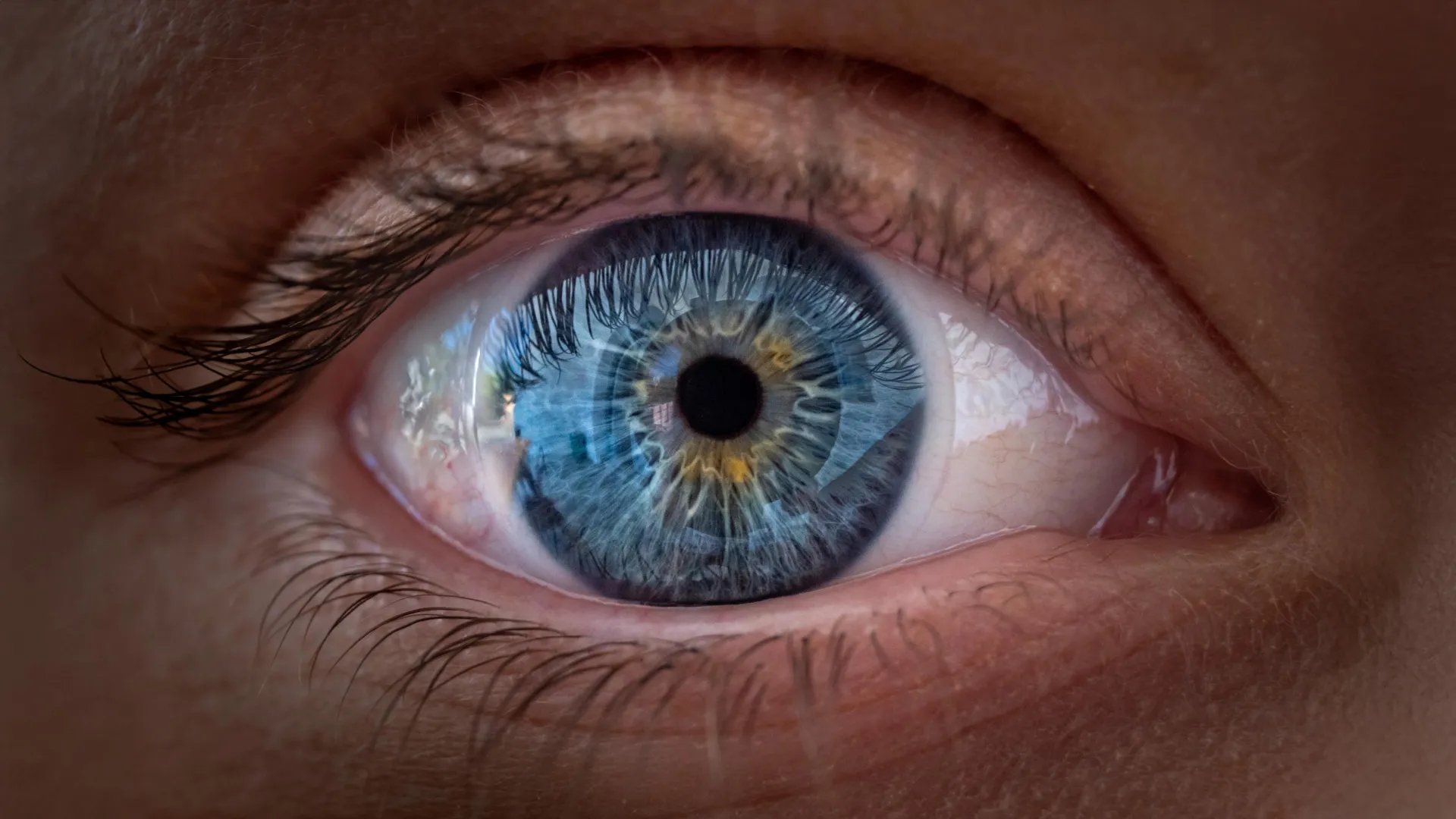
Scientists at Johns Hopkins University have found that vitamin A derivatives and thyroid hormones work together to shape the human fovea, the retinal region responsible for our sharpest central vision. Their findings suggest that some blue-sensitive cone cells present early in fetal development may later transform into red- and green-sensitive cones, challenging a model that has influenced vision research for nearly 30 years.
Recreating the developing human retina
The study, published in Proceedings of the National Academy of Sciences, examines changes that occur in the retina before birth. The results could improve scientists’ understanding of how the human eye develops and may eventually inform research into treatments for macular degeneration, glaucoma, and other causes of vision loss.
“This is an important step toward understanding the inner workings of the central retina, a critical part of the eye and the first to fail in people with macular degeneration,” said Robert J. Johnston Jr., an associate professor of biology at Johns Hopkins University who led the study. “By understanding this region better and developing organoids that mimic its function, we hope to one day grow and transplant these tissues to restore vision.”
To study how human vision forms, Johnston and his colleagues used miniature versions of human retinal tissue known as organoids. The researchers have helped develop these experimental models over the past several years.
The organoids are small clusters of cells grown from fetal cells that reproduce important features of developing human tissue. By observing them over several months, the scientists could track changes that would be difficult to study during pregnancy.
The researchers focused on the fovea, a small area at the center of the retina. The retina is a light-sensitive layer at the back of the eye that converts incoming light into signals the brain can interpret. The fovea is specialized for detailed central vision, allowing people to read, recognize faces, and distinguish fine details.
Although the fovea occupies only a small part of the retina, researchers say it accounts for about 50% of human visual perception.
The cellular basis of sharp central vision
The fovea’s function depends in part on specialized light-detecting cells called cones. Rod cells are particularly important for vision in dim light, while cones support daytime vision and color perception.
Humans have three types of cones, each most sensitive to a different range of wavelengths. They are commonly described as blue, green, and red cones. The brain compares the signals from these cells to distinguish among a wide range of colors.
Cones are not distributed evenly across the retina. The fovea contains red and green cones but lacks the blue cones found in other retinal regions. This precise arrangement is essential to the clear vision produced by the center of the eye.
Scientists have struggled for decades to determine how this pattern develops. Commonly studied animals, including mice and fish, do not have the same retinal structures as humans, limiting the usefulness of traditional animal models for investigating the process.
Human retinal organoids have provided a new way to examine this long-standing question.
How vitamin A and thyroid hormones shape the fovea
The researchers found evidence that the foveal cone pattern develops through two coordinated processes. These processes help determine which cone types form in the central retina and how some cells change as development continues.
Between 10 and 12 weeks of fetal development, a small number of blue cones appear in the fovea. By week 14, however, these cells appear to have transformed into red and green cones.
The team identified two chemical influences involved in this process. The first is retinoic acid, a molecule produced from vitamin A. Retinoic acid helps regulate cell development and specialization throughout the body. In the developing retina, the breakdown of retinoic acid limits the formation of blue cones in the central region.
The second influence is thyroid hormone. Although thyroid hormones are best known for regulating metabolism, they also play important roles in growth and development. In the developing fovea, the hormone appears to encourage the remaining blue cones to transform into red and green cones.
“First, retinoic acid helps set the pattern. Second, thyroid hormone is responsible for transforming the remaining cells,” Johnston said. “This is very important because if you have blue cones there, your vision will be impaired.”
The findings concern the role of vitamin A signaling in prenatal eye development. They do not establish that taking vitamin A supplements can improve clear vision in adults.
A challenge to a decades-old theory
The results offer an alternative to a model that has shaped vision research for roughly 30 years. Under that long-standing explanation, blue cones appeared in the fovea and later moved away as the retina developed. Scientists generally assumed that once a cone became a particular type, its identity remained fixed.
The new findings suggest that at least some blue cones may not simply change position. Instead, they may change identity and become red or green cones as the eye develops.
“The leading model in this field about 30 years ago was that some blue cones that you get in that area somehow move out of the way, determine what these cells will become in the future, and remain this type of cell forever,” Johnston said. “We still can’t completely rule it out, but our data support a different model: These cells actually transform over time, which is really surprising.”
The researchers have not ruled out the possibility that some cells migrate. Nevertheless, their observations support the idea that developing cells in this region are more flexible than previously recognized.
This possibility could change how scientists investigate the formation of human color vision and the development of the retina’s most specialized regions.
Potential implications for restoring vision
The findings may help explain not only how healthy eyes develop but also how researchers might eventually repair retinas damaged by disease.
One possible application involves macular degeneration, a major cause of central vision loss, particularly among older adults. The disease damages the macula, the part of the retina responsible for detailed central vision, and can interfere with reading, driving, and recognizing faces. Treatments can slow or manage the progression of some forms of the disease, but there is currently no cure.
A better understanding of how the retina produces specialized light-sensitive cells could eventually help scientists generate healthy replacement cells in the laboratory.
Johnston’s team is working to improve retinal organoids so they more closely reproduce the structure and function of the human eye. The long-term aim is to gain precise control over populations of photoreceptors, the cells that detect light, with the possibility of transplanting them into damaged retinas.
Study author Katarzyna Hussey, a former doctoral student in Johnston’s laboratory, said the approach could open new avenues for restoring vision. Hussey is now a molecular cell biologist at CiRC Biosciences, a cell therapy company in Chicago.
“The goal of using this organoid technology is to eventually create a nearly custom-tailored population of photoreceptors. A big possibility is cell replacement therapy, which introduces healthy cells that could reintegrate into the eye and potentially restore lost vision,” Hussey said.
For such a treatment to work, transplanted cells would need to survive, connect correctly with the eye’s existing circuitry, and function safely. Hussey emphasized that the technology remains far from routine clinical use and requires extensive development and testing.
“These are very long-term experiments, and of course we need to do some optimization for safety and efficacy studies before we move into the clinic. But it’s a viable journey.”
For now, the discovery provides a clearer account of how the human eye develops its ability to see fine details. By revealing how vitamin A signaling and thyroid hormones help shape the fovea, the research offers new insight into early vision development and a possible foundation for future efforts to restore sight after retinal disease.
Source: www.sciencedaily.com