


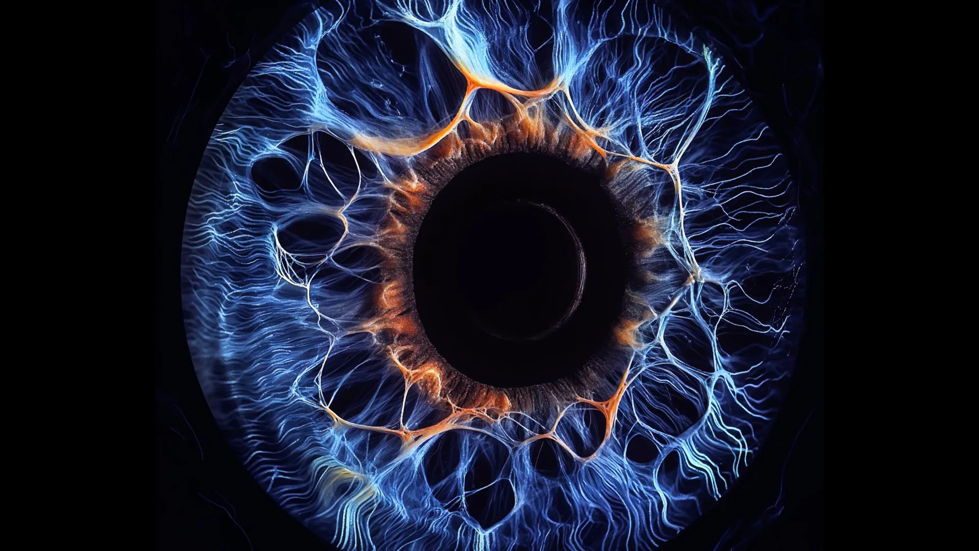
New insights from Yale School of Medicine (YSM) reveal that the human eye processes visual information in ways far more intricate than previously understood. This groundbreaking research challenges established beliefs about how visual signals traverse the retina and provides new explanations for our ability to detect faint objects and see in low-light conditions.
Our visual system rapidly analyzes various elements of a scene, including color, contrast, movement, and shape. This process, known as parallel visual processing, enables the brain to interpret complex images almost instantaneously by sending different types of information along distinct pathways.
Traditionally, researchers believed that these pathways operated independently as visual signals traveled from the retina to the brain. However, a 2016 study indicated that neuron connections are intimately linked through hidden electrical pathways. Researchers assert that this interconnectedness could enhance weak visual signals before they proceed deeper into the visual system.
“We found that while distinct channels offer unique functionalities, they are interconnected by underlying electrical circuits,” states Dr. Yao Xue, a postdoctoral fellow in YSM’s Department of Ophthalmology and Visual Sciences and the study’s lead author.
The Role of Bipolar Cells in the Visual Network
Vision begins when the retina’s rods and cones detect light. These specialized cells relay information to bipolar cells, a type of neuron. At this initial stage, visual data is processed in parallel channels that differentiate aspects such as daylight clarity, night vision, color, contrast, and shape.
Upon closer examination of synapses—the microscopic junctions where bipolar cells communicate—researchers uncovered surprising results. Channels anticipated to be isolated were, in fact, sharing information with one another.
Neurons communicate primarily through two types of synapses: chemical and electrical. Chemical synapses transmit information using neurotransmitters, while electrical synapses, or gap junctions, facilitate direct signal transfer. Contrary to previous beliefs, bipolar cells were found to utilize both methods for communication.
New research identifies that electrical synapses link most of these isolated information channels in both mouse and human retinas. When researchers stimulated a single bipolar cell electrically, the response extended far beyond the expected pathway, revealing a broad, ‘cloud-like’ pattern of activity indicative of extensive communication among different bipolar cell types.
“When we stimulated one bipolar cell, multiple bipolar cells simultaneously released neurotransmitters,” commented lead researcher Z. Jimmy Chou, Ph.D., Marvin L. Sears Professor of Ophthalmology and Visual Sciences.
The study also identified a specific type of bipolar cell, termed BC6, pivotal in coordinating this elaborate network. Signals originating from BC6 propagated through various visual pathways in a systematic and organized manner.
“People previously viewed different bipolar cell types as relatively autonomous,” Zhou notes. “However, we discovered that all cell types contribute drivers that create this hierarchical network.”
Scientists assert that this combination of specialized pathways and electrical communication endows the retina with a dual advantage. Distinct channels enable concentration on specific visual attributes, while interconnected channels collaborate to enhance weak signals.
“When signals are faint and split across multiple channels, minimal processing occurs in each,” explains Seunghoon Lee, Ph.D., a researcher in YSM’s Department of Ophthalmology and Visual Sciences and a co-author of the study. “This integration proves particularly beneficial for detecting low-contrast or small object signals.”
“Cells don’t cooperate randomly,” Xue added. “There exists a leader—BC6—that orchestrates the relay of signals to downstream targets.”
Innovative Mapping of Retinal Signals
To elucidate these communication networks, the research team employed advanced techniques. They utilized cutting-edge imaging technologies to monitor how bipolar cells released neurotransmitters while simultaneously stimulating individual cells and recording adjacent responses.
Historically, studying bipolar cells has been challenging due to their deep retinal placement. Earlier experiments required slicing the retina, which could disrupt the natural circuitry under investigation.
This study marks a breakthrough, as the Yale team effectively used a dual patch clamp technique on an intact mouse retina. Researchers expertly stimulated specific bipolar types and recorded how neighboring cells reacted.
“No other laboratory globally has achieved systematic recording of this nature,” Zhou asserts. “This work stands as an innovative achievement from Yao Xue’s doctoral research, merging unique approaches with exceptional electrophysiological techniques.”
The team replicated these experiments using intact human retina sourced from the Department of Pathology’s Legacy Tissue Donation Program, making this the first study of its kind on unaltered human retinas.
Implications of the Discovery
Given that the retina is part of the central nervous system, these findings possess potential implications beyond visual perception. Insights gained from understanding retinal circuitry may reveal new perspectives on how other neural networks in the brain operate.
The research could also enhance understanding of retinal diseases such as macular degeneration, glaucoma, and congenital night blindness.
Furthermore, the study underscores the significance of curiosity-driven science. Rather than leading with a predefined hypothesis, this investigation unveiled an unknown mechanism that shifts how scientists conceptualize visual processing.
“Our experiments began without a specific hypothesis, allowing us to uncover a fundamental processing mechanism in the visual system,” remarks Lee. “This serves as a crucial reminder of the importance of curiosity-driven research in fostering scientific discovery.”
Source: www.sciencedaily.com