


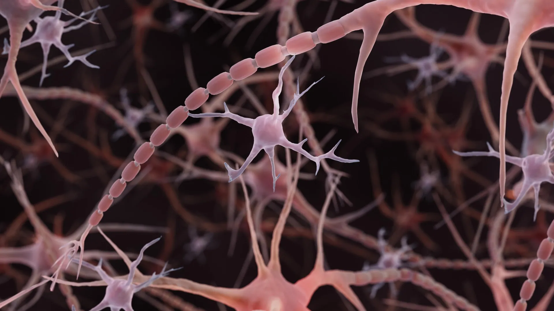
Beaded Axons May Change How Brain Signals Travel
The brain’s fine wiring may require a different kind of textbook illustration. Instead of the smooth, thin tubes typically shown extending from nerve cells, some axons resemble strings of tiny pearls. These repeating bulges are more than decorative details: their shape appears to influence how electrical messages move through the brain.
Researchers at Johns Hopkins School of Medicine highlighted this unexpected architecture in a study published online in Nature Neuroscience. Using mouse neurons, they found evidence that familiar diagrams of brain cell anatomy may overlook important features of the extensions that carry signals.
Related research later identified pearlescent axons in human brain tissue, extending the observations beyond mice. Together, the findings raise an important question: how does the physical structure of the brain’s wiring help regulate communication?
“Understanding the structure of axons is important for understanding brain cell signaling,” said Shigeki Watanabe, Ph.D., associate professor of cell biology and neuroscience at the Johns Hopkins University School of Medicine, in a university presentation in 2024. “Axons are the cables that connect brain tissue and enable learning, memory, and other functions.”
A new view of brain cell axons
Axons are long extensions that allow neurons to transmit electrical signals to other cells. Traditional diagrams show relatively uniform tubes interrupted by occasional bulges called synaptic varicosities. These structures contain groups of neurotransmitters that help transmit signals to other brain cells.
Beaded axons themselves are not uncommon. Scientists have observed substantial swelling in dying neurons and in neurodegenerative conditions such as Parkinson’s disease. In these situations, bead formation can damage the cell membrane and the internal structures that help maintain axonal shape.
What was striking about the Johns Hopkins study was the presence of much smaller, repeated swellings in axons examined under conditions designed to preserve normal structure. The researchers called these areas “nonsynaptic varicosities.” Unlike familiar bulges associated with communication sites, these structures were not synapses.
The distinction is important. The study did not suggest that all beaded axons are damaged. Instead, it identified a nanoscale, pearl-like morphology that may be part of the normal structure of some axons.
Clues from insects and the axon’s internal skeleton
Watanabe’s interest began with the observation that pearls repeat along the axons of insects. A conversation with Swiss scientist Dr. Graham Knott prompted him to investigate the phenomenon further. One possible explanation involved the axon’s internal skeleton, a supporting network of proteins.
In a 2012 study, a Harvard University team reported that skeletal components are repeated within axons. Watanabe and Knott wondered whether destroying this framework would cause the pearls to disappear. Jacqueline Griswold, a Johns Hopkins graduate student and lead author of the study, tested the idea. The pearls remained.
This result directed the researchers’ attention to the physical properties of axons. Watanabe and Griswold joined Padmini Rangamani, a theoretical biophysicist and professor of pharmacology at the University of California, San Diego School of Medicine, to investigate how surrounding membranes shape axons.
Freezing brain cells reveals hidden axon structures
Studying these structures requires imaging features much narrower than a human hair. Johns Hopkins described the axons as approximately one-hundredth the width of a human hair. The researchers used high-pressure cryo-electron microscopy to preserve their delicate shape before examining them with an electron beam.
The preparation method was important. Standard electron microscopy typically involves chemically fixing and dehydrating tissue, steps that can alter the structures scientists are trying to observe. Freezing helps preserve the tissue’s original shape.
“To see nanoscale structures with a standard electron microscope, you fix the tissue and dehydrate it, but when you freeze it, it retains its shape. It’s like freezing grapes instead of dehydrating them into raisins,” Watanabe said.
The researchers examined neurons grown in the laboratory, neurons from adult mice, and neurons from mouse fetuses. Their axons were unmyelinated, meaning they lacked the insulating myelin covering found around many axons. Across tens of thousands of tissue images, researchers repeatedly observed the same pearl-like appearance.
They also observed pearl formation in high-resolution images of living neurons, providing evidence that the pattern was not simply caused by freezing the samples.
“These discoveries challenge a century of understanding of axonal structure,” Watanabe said.
How membrane physics shapes axons
To explain the pattern, the researchers built a mathematical model of the membrane surrounding axons. The relatively simple mechanical model reproduced key characteristics of the pearls, suggesting that physical forces may help determine axon shape without requiring a rigid internal mold.
Experiments supported this interpretation. Increasing the sugar concentration in the solution surrounding the axon reduced the size of the swollen regions. In the model, increasing membrane tension also reduced the size of the membrane bulges.
Cholesterol provided another way to alter membrane behavior. When cholesterol was removed, the membrane became less rigid and more fluid. This changed the pearl structure in both the model and mouse neurons and slowed electrical signal transmission.
Watanabe compared the effect to traffic moving through a wider space: a larger area within the axon allows ions to pass through more easily. However, larger pearls did not always produce faster signals. The dimensions of both the bulge and its narrow connection were important.
Electrical activity can change axon shape
The researchers also tested whether neural activity could alter axon structure. After high-frequency electrical stimulation, the pearl-like regions became an average of 8% longer and 17% wider. The enlargement lasted for at least 30 minutes.
The published study reported that electrical signals slowed after the stimulation, and the effect lasted for at least an hour. When cholesterol was removed beforehand, some structural changes still occurred. However, the pearls remained smaller than those in untreated cells, and researchers did not detect a similar significant slowing of signal transmission.
The findings suggest that axons are not simply fixed cables. Their physical structures can respond to activity and help regulate how messages travel through neurons.
Related research finds pearl-shaped axons in human brain tissue
When the initial findings were announced in 2024, the research team was planning to examine axonal “arms” in human brain tissue obtained with permission from people undergoing brain surgery or affected by neurodegenerative diseases.
A related study led by Chelsy R. Eddings and colleagues, including Watanabe, later reported pearlescent axons in human cortical tissue obtained during epilepsy surgery. The study was posted online and published in Neuron in the November 24, 2025, and February 4, 2026, issues.
Researchers combined electrical stimulation with flash-freezing to capture small membrane changes in mouse and human brain slices. They found evidence of ultrafast endocytosis alongside pearly axons. Endocytosis is the process by which nerve endings rapidly retrieve membrane after releasing a chemical signal.
Although these observations extend the evidence to human tissue, they do not establish that all axons have the same shape. Researchers also have not shown that pearling affects signal transmission in the same way throughout the human brain. Human studies have primarily investigated membrane recycling at synapses.
Researchers continue investigating what controls axon shape
The relationship between axon structure and function is being explored by Watanabe and Rangamani, as described in a 2024 National Institute of Mental Health project. The work will support computational models of the physical properties of neurons and examine how incoming signals affect their axons. Researchers will then test the models in mouse tissue and cell cultures.
Other research is exploring additional ways to investigate the dimensions of these fine neural connections. In a study published in PLOS Biology on July 17, 2026, researchers at the University of Edinburgh screened 880 compounds for their effects on axon diameter using automated imaging of live zebrafish. They identified 33 initial hits and confirmed that compounds affecting specific chemical-signaling pathways can increase axon width.
Although the zebrafish study does not directly replicate research on pearl formation, it provides another tool for investigating how axon size is controlled.
For the Johns Hopkins team, an important question remains: how are the structures found in functioning axons related to the damaging beads associated with neurological diseases? Understanding the difference could help researchers distinguish normal changes in neural wiring from signs of cellular failure.
Source: www.sciencedaily.com