


Alkalineptosis and Anastasis: How Cells Die—and Sometimes Recover
Cells in Daolin Tan’s petri dish were dead. Traces of life were falling from the edge of the culture, and the cells were no longer reproducing. They were failures.
That was exactly what Tan and his colleagues wanted. Cell biologists were searching for chemicals that could poison cancer cells.
The secret battle between your body’s cells may save your life
But Tan and his colleagues faced a post-mortem mystery: what had killed the cells? They investigated several known causes of cell death, but none appeared to be responsible.
Eventually, the researchers found that the internal pH of the dying cells was abnormally high. The cells had died because they were too alkaline.
The research team named this phenomenon alkalineptosis.1 The study expanded scientists’ understanding of how cells die and how pH controls cell fate. It also suggested a potentially new way to kill cancer cells, says Tan, who conducted the research while at the University of Pittsburgh in Pennsylvania and now works at the University of Texas Southwestern Medical Center in Dallas.
Tan’s experience has been repeated many times in the twenty-first century. Since 1999, scientists have reported around 20 new types of cell death, and the number continues to grow.
Scientists are discovering new forms of cell death
For decades, researchers believed that cells died in two main ways. Death could occur accidentally, when cells were crushed, sliced or otherwise destroyed. This process is called necrosis.
The other classic pathway is apoptosis, a highly regulated and orderly form of cell death. During apoptosis, cells release tiny membrane-bound structures called vesicles, which are cleared away by the immune system.
For a long time, these two options dominated the field, perhaps because cell death is difficult to study. Scott Dixon, a cell biologist at Stanford University in California, jokes that when a cell-death experiment succeeds, the experimental material disappears.
Modern technologies—including gene editing, high-resolution imaging and a variety of “omics” tools—have revealed many additional pathways, says cell biologist Xinbin Hu of Xijing Hospital in Shaanxi Province, China.
These include inflammatory pyroptosis, explosive immune-activating necroptosis and ferroptosis, in which iron contributes to membrane damage. At least four new types of cell death were defined in 2025 alone.
This spring, scientists studying flatworms reported another phenomenon called raptosis. In this process, certain cells explode and spray toxins onto invading bacteria.2 Cells may also possess mechanisms that delay death—or even allow them to pull back from the brink and survive.

Cells called raptoblasts, discovered this year, can explode and release immune substances that kill invaders.
Credit: Dr. Elijah Sultan
How cells die matters, Tan says. Dying cells release signals and substances that affect neighboring cells, shape tissues and influence immune responses and disease.
For example, after being invaded by a pathogen, a cell may undergo programmed death. Once it dies, however, its contents can leak out and call immune cells to fight a wider infection. Understanding how cells die—and why they sometimes fail to die when they should—could therefore clarify the biology of development, metabolism and immunity.
Scientists are eager to connect newly discovered death pathways to disease. Potential medical applications include killing cancer cells, calming inflammatory diseases and rescuing dying cells in the heart and brain.
The discovery of these pathways highlights how much remains unknown about basic biology, Tan says. “Cell death is not simply the end of a cell’s lifespan.”
Why cells have so many ways to die
Every day, billions of cells in the human body are removed. When damaged, worn out or no longer needed, they wither, harden or explode.
“Every cell in a living thing needs to know when, how and where to die,” Hu says. “With so many different cells playing unique roles in life, it’s no wonder that different forms of cell death are so needed.”
Excess amounts of certain compounds can also kill cells, and the resulting form of cell death depends on the cause. Researchers have described cell death caused by excess copper.3 In 2022, another study examined death caused by excessive sodium.4
These findings reveal the limits of cell survival, says Qing Chong, a biochemist at Shanghai Jiao Tong University School of Medicine in China who led the study on sodium-induced cell death.

Cells have a menu of self-induced death programs, including apoptosis and pyroptosis. If everything works properly, these pathways benefit the organism.
During development, for example, cells that create webbed fingers in human fetuses undergo apoptosis after their work is complete. Cells with limited lifespans, such as those lining the intestines, are replaced by new cells. Some skin cells undergo a form of death called keratinization, leaving behind dead cells that form the body’s protective outer layer.
Infected cells often sacrifice themselves to prevent viruses and bacteria from multiplying. Ana Garcia-Saez, a biophysicist at the Max Planck Institute for Biophysics in Frankfurt, Germany, suggests that infection may have helped drive the evolution of some cell-death pathways.
Infected cells may have needed backup options to ensure that they could eliminate themselves. Some viruses can keep host cells alive by blocking individual death pathways. Even if a cell can switch to another program, such as pyroptosis or necroptosis, the virus may be thwarted.
“It’s like a competition with pathogens,” Garcia-Saez says.
When cells delay death—or come back to life
Scientists have also discovered that cell death is more flexible than previously thought. Cells do not always move directly from a death trigger to complete destruction. They may linger in a damaged state or even survive.
Consider intestinal epithelial cells. Dying cells normally remove themselves from the intestinal lining and help with their own disposal. This process takes time, so the cells must remain alive long enough to complete it.
A 2022 study by Edward Miao, an immunologist at Duke University in Durham, North Carolina, and Kengo Nozaki, then a postdoctoral fellow and now an immunologist at the Medical University of South Carolina in Charleston, examined the process.5
The researchers studied mice lacking caspase-7, an enzyme that promotes cell death, and infected them with Salmonella, bacteria that cause food poisoning. The infected intestinal epithelial cells began to die. Without caspase-7, however, the cells became stuck in the intestinal lining, causing additional inflammation and tissue damage.
Nozaki and Miao found that the dying cells developed holes in their membranes. Caspase-7 normally activates membrane repair, allowing the cells to survive long enough to complete a clean exit from the intestinal lining.
In a 2023 review, Nozaki and Miao proposed that many cells have a “bucket list” of tasks to complete before they expire.6
Some immune cells use these final functions to protect the body from pathogens. Macrophages infected with bacteria can die by pyroptosis, releasing distress signals and arranging their membranes to trap pathogens on their dead bodies.7
Neutrophils, which live for only a few hours to a few days, can also behave aggressively before they die. When they encounter bacteria, their DNA unravels and the nuclear membrane dissolves. The cell may then rupture, releasing a net of its own genetic material that traps invading organisms.

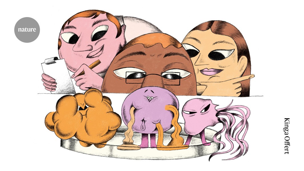
Illustration: Kinga Ofert
Petr Broz, an immunologist at the University of Lausanne in Switzerland, calls the bucket-list idea “a really great concept” but says it is difficult to prove experimentally.
Once cell death begins, it is not necessarily inevitable, says Ho Lam Tan, a cell biologist at Johns Hopkins University School of Medicine in Baltimore, Maryland.
Tan and his sister, Ho Man Tan, a tumor biologist and postdoctoral fellow in another Johns Hopkins laboratory, observed this in 2007 while studying apoptosis at the Chinese University of Hong Kong. At the end of an experiment, they noticed that small, struggling cells remained in the culture dish.
They washed out the cell-death inducer and incubated the shrunken cells in fresh medium. The next morning, many of the cells had recovered.

Two human white blood cells. One undergoes apoptosis, a form of programmed cell death.
Credit: Dr. Gopal Murti/Science Photo Library
The cells appeared to revive themselves by activating several repair pathways. The researchers called this process anastasis, a Greek word meaning “resurrection.” Their findings challenged the common belief that apoptosis is irreversible once it begins.
The brothers submitted their findings to 11 journals before editors were convinced by a video showing the cells reviving. Molecular Biology of the Cell eventually published the work.8
Since then, researchers have observed the reversal of ferroptosis and necroptosis and expect that other forms of cell death may also be reversible.
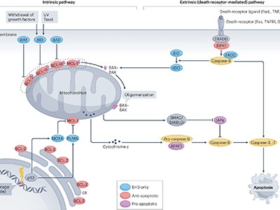
Guide to cell death pathways
The sometimes reversible nature of cell death suggests that the point of no return comes much later than scientists once thought. Broz and Garcia-Saez both speculate that disease develops when a cell’s mitochondrial boundaries are irreparably disrupted and its metabolism disappears.
“At some point, when you die, you die,” Garcia-Saez says.
Source: www.nature.com