


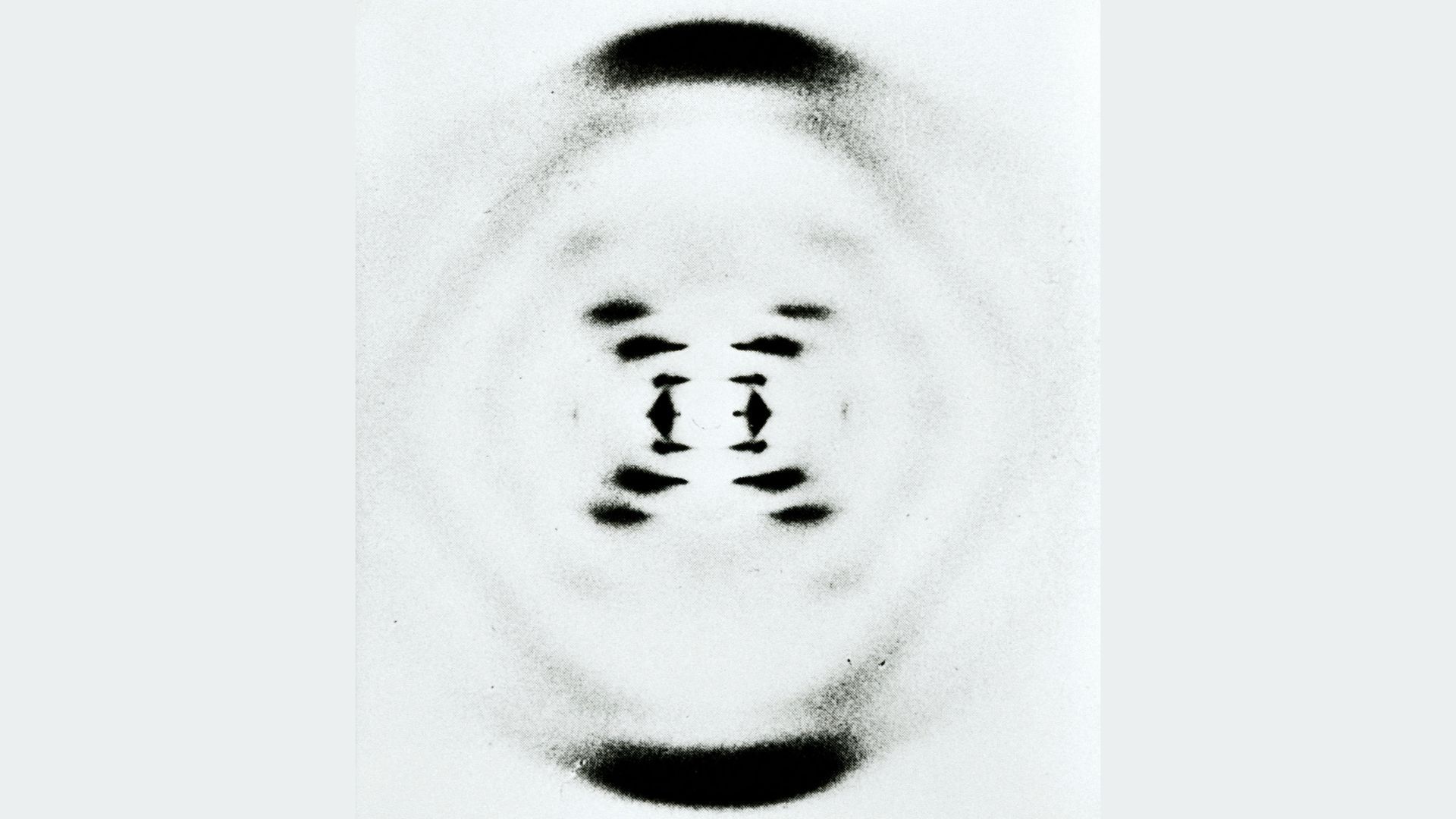
Rosalind Franklin Knew Photo 51 Revealed DNA’s Helical Structure, New Study Suggests
Rosalind Franklin likely understood that her famous X-ray image of DNA contained crucial evidence of the molecule’s helical structure, according to a new study of archival materials.
Known as Photo 51, the image was taken in May 1952 and later published as evidence supporting DNA’s double-helix structure. The new research challenges the idea that Photo 51 was merely an unimportant experimental image that Franklin failed to interpret.
Instead, the archives suggest that Franklin deliberately produced Photo 51 as a polished image suitable for publication. She began taking it on the same day she viewed an earlier image, Photo 49, which showed the same DNA sample.
“She really wanted to get the polished version, so she took it,” said study co-author Alistair Sponsel, a historian of science at the Philadelphia Institute for the History of Science. Taking a better photograph of the same sample was “a sign that she knew she had something really important,” Sponsel told Live Science.
What Photo 51 revealed about DNA
Photo 51 was later shared with Francis Crick and James Watson by Maurice Wilkins. Watson described the image in his 1968 autobiography, The Double Helix: A Personal Account of the Discovery of the Structure of DNA, calling it an “aha” moment.
“The reflection of the black cross that dominated the photograph could only have arisen from the helical structure,” Watson wrote.
Image credit: Donaldson Collection via Getty Images
Franklin has sometimes been portrayed as highly skilled in X-ray crystallography but unable to interpret the significance of her own data. However, the archival evidence presents a more complicated picture.
The new study, published Monday, October 5, in the Biological History Journal, indicates that Franklin’s notes included an early description of a possible spiral shape in Photo 49. She later created the more refined Photo 51 to show the findings more clearly.
Why Franklin created two DNA images
“In Franklin’s mind, these were two different types of scientific images,” Sponsel said. Photo 49 was an experimental image that revealed new data, while Photo 51 was an exhibit intended to communicate those findings as clearly and vividly as possible.
Sponsel conducted the study with molecular biophysicist Brian Sutton, who earned his PhD from King’s College London. Their research began after the Institute for the History of Science acquired archival materials from Franklin and her graduate student Raymond Gosling.
The researchers also examined archives at King’s College London, where Franklin was working when she took the famous images.
Franklin’s research on DNA structures
Franklin’s lack of public notes about Photo 51 has been used to suggest that she missed a critical discovery. But Sponsel said her journal entry for Photo 49 described a possible spiral shape and contained significant structural information. Photo 51 followed as a more sophisticated presentation of the same findings.
Franklin’s scientific goal, outlined in a report from February 1952, was to understand two distinct forms of DNA known at the time as A and B. Structure B, shown in Photos 49 and 51, is the form that naturally occurs in cells and has a double-helix structure. Structure A is the form DNA takes when dehydrated in the laboratory.
Structure A appeared to contain more data, so Franklin chose to characterize it before studying Structure B in depth. While she was doing that work, Wilkins showed Watson an image of Structure B. Watson and Crick then worked with Wilkins to build a detailed model of DNA’s double helix.
In 1962, Watson, Crick and Wilkins shared the Nobel Prize in Physiology or Medicine for their work on DNA’s structure. Franklin had died of ovarian cancer in 1958 and was therefore ineligible for the award. The importance of her contributions became more widely recognized in the years that followed.
Editor’s note: This article was updated at 3:23 p.m. ET to clarify that Rosalind Franklin likely recognized DNA’s helical structure, but not necessarily the complete double-helix model. The image at the top is another photograph taken by Franklin, not Photo 51.
Source: www.livescience.com