


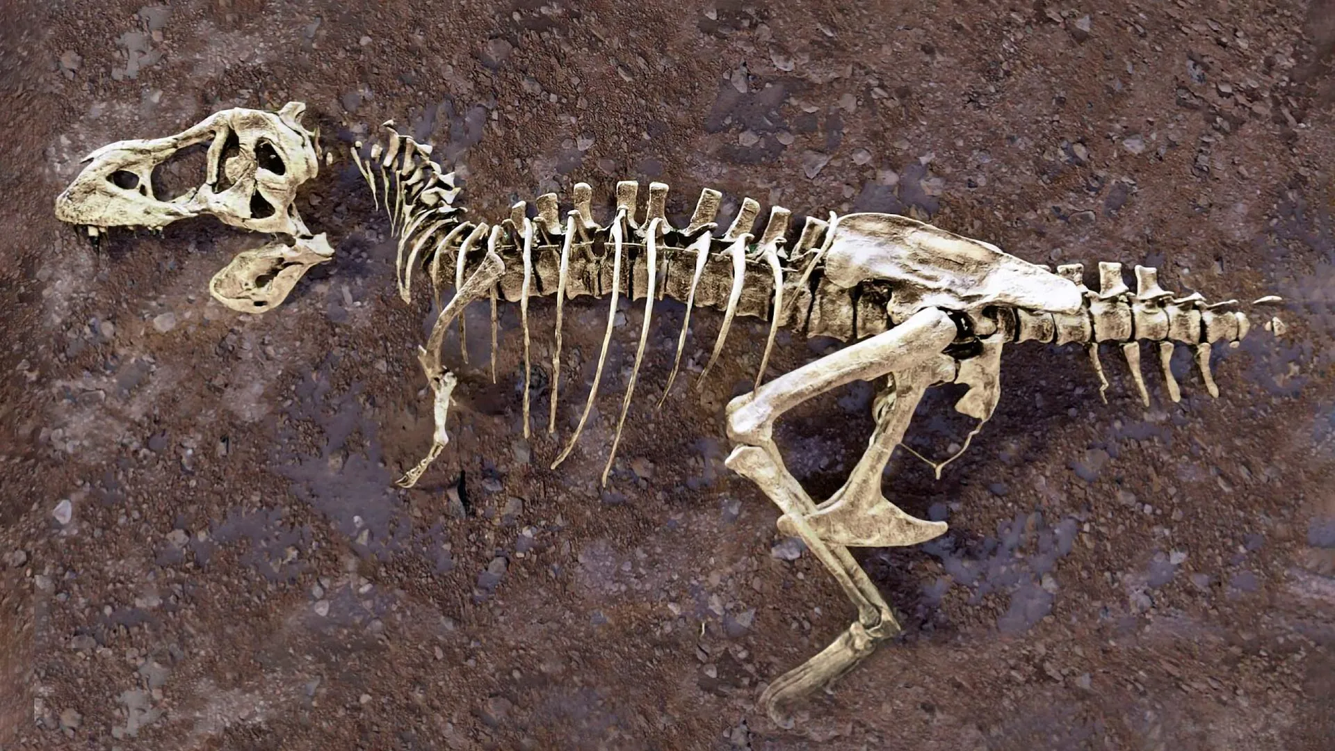
Scotty the Tyrannosaurus rex’s Healing Rib Fractures Reveal Rare Fossilized Blood Vessels
Neutron imaging of Scotty, one of the most notable Tyrannosaurus rex skeletons ever discovered, is giving researchers a rare look at dinosaur biology 66 million years ago. Inside the fossilized ribs, scientists found evidence of injuries that were still healing when the dinosaur died—including a vast network of calcified blood vessels rarely preserved in the fossil record.
Neutron imaging reveals Scotty’s fossilized blood vessels
Scientists at the U.S. Department of Energy’s (DOE) Oak Ridge National Laboratory (ORNL) used neutron imaging to examine Scotty’s fossil from the inside. The non-destructive technique produces detailed 3D images while preserving the fossil and its soft-tissue structures.
“It’s like winning the lottery,” said Mauricio Balbi, a physics professor at the University of Regina in Saskatchewan, Canada. “Scotty’s ribs contain a vast network of calcified blood vessels never before observed in fossils.”
Fossilized blood vessels preserve evidence of injury and healing
Soft tissues such as blood vessels are usually lost during decomposition long before fossilization is complete. Scotty’s ribs are a rare exception, preserving delicate structures that provide a glimpse into what was happening inside the dinosaur as its injuries healed.
After a rib broke, iron-rich blood entered the injury site and new blood vessels formed as part of the healing process. Scotty died before the fractures had fully healed. The dinosaur later ended up in salty swamps, where environmental conditions helped slow decay and preserve the fragile network of blood vessels.
“Every fossil is a small snapshot of the past,” said Gerrit Mitchell, a doctoral student in physics who is leading the project under Balbi’s guidance.
Scotty’s remains were discovered by a team from the Royal Saskatchewan Museum in Saskatchewan’s Frenchman River Valley, one of the richest dinosaur fossil sites in North America. Rocks in the region preserve important records of dinosaur life shortly before the mass extinction that ended the era of non-avian dinosaurs. Researchers are also studying fossilized amber, dinosaur scales, and bones from other dinosaurs found there.
“By piecing together clues, we can understand the past and how things will develop in the future,” said Marcella Berg, an assistant professor of physics at the University of Tokyo and a former ORNL postdoctoral researcher.
Neutron and X-ray imaging reveal a dinosaur’s medical history
Neutron and X-ray imaging provide complementary ways to examine the inside of fossils and other materials. Neutrons are particularly useful for detecting lighter elements on the periodic table, especially hydrogen, while X-rays are highly effective at revealing heavier elements.
The difference is similar to the contrast between MRI, which can highlight soft tissues such as muscles, and X-rays, which are especially useful for viewing dense structures such as bones. Researchers select different neutron and X-ray methods depending on the material and features they want to study.
The research dates back to 2020, when Mitchell, then an undergraduate at the University of Regina, detected traces of blood vessels inside Scotty’s ribs. The Canadian Light Source was the first facility to use micro-CT scanning—a non-invasive X-ray imaging method—to examine the fossil. Those scans revealed fossilized soft tissue on the cut surfaces of the ribs.
As the investigation expanded, researchers combined microscopy with other X-ray techniques, including Canadian Light Source synchrotron radiation. Together, these methods allowed scientists to study both wound healing and preserved fossil tissue at the cellular level.
After X-ray images revealed evidence of fossilized blood vessels, the researchers turned to neutron imaging to search for additional clues in the blood vessels and other preserved soft tissues.
Powerful neutron beams examine Scotty’s ribs without damaging them
In April 2026, researchers used the Multimodal Advanced Radiography Station (MARS) at ORNL’s High Flux Isotope Reactor (HFIR) and the Neutron Science Virtual Environment (VENUS) instrument at the DOE’s Spallation Neutron Source (SNS).
Neutron imaging allowed the team to confirm previous observations, examine large bones such as Scotty’s ribs without damaging them, and obtain additional image contrast that complemented information gathered through other methods.
“Neutrons not only confirm what we discovered with synchrotron radiation techniques that led to the discovery of Scotty’s rib vessels, but they also prove to be an extremely valuable addition to our current work seeking to preserve soft tissue in fossils,” Berg said. “This provides incredible detail to better understand these properties without affecting the sample.”
MARS produces cold neutrons and is particularly effective at highlighting features associated with soft tissue, hydrogen-rich regions, and subtle differences within a specimen. Researchers used the instrument to generate high-resolution images of small bone, amber, and scale fossils.
In contrast, VENUS produces high-energy neutrons that can penetrate deep into large objects and create detailed 3D images. Researchers used VENUS to examine large bones, including Scotty’s ribs.
A new way to search for hidden biology in fossils
As neutrons pass through a sample, they interact with atoms throughout the material and are particularly sensitive to hydrogen atoms. The resulting information can be transformed into images. Because neutrons behave differently from X-rays, they can reveal features that are difficult or impossible to detect with other imaging techniques.
“People tend to think of neutrons as a tool for studying batteries and advanced materials, but they are equally transformative for answering questions about ancient life,” said Haseena Billhew, VENUS’ chief instrument scientist.
Researchers will continue studying data collected by VENUS and MARS while expanding their approach to additional fossils. They also plan to compare patterns of injury and healing among different species.
By combining neutron imaging with X-ray techniques, scientists hope to investigate pathologies preserved in fossils and compare the ancient conditions they reveal with differences observed in modern species.
“There are more fossils in our collection than you think, hiding secrets from millions of years ago,” Mitchell said. “By putting them into a synchrotron or neutron source, we can make new discoveries about ancient life that have never been made before.”
Source: www.sciencedaily.com