


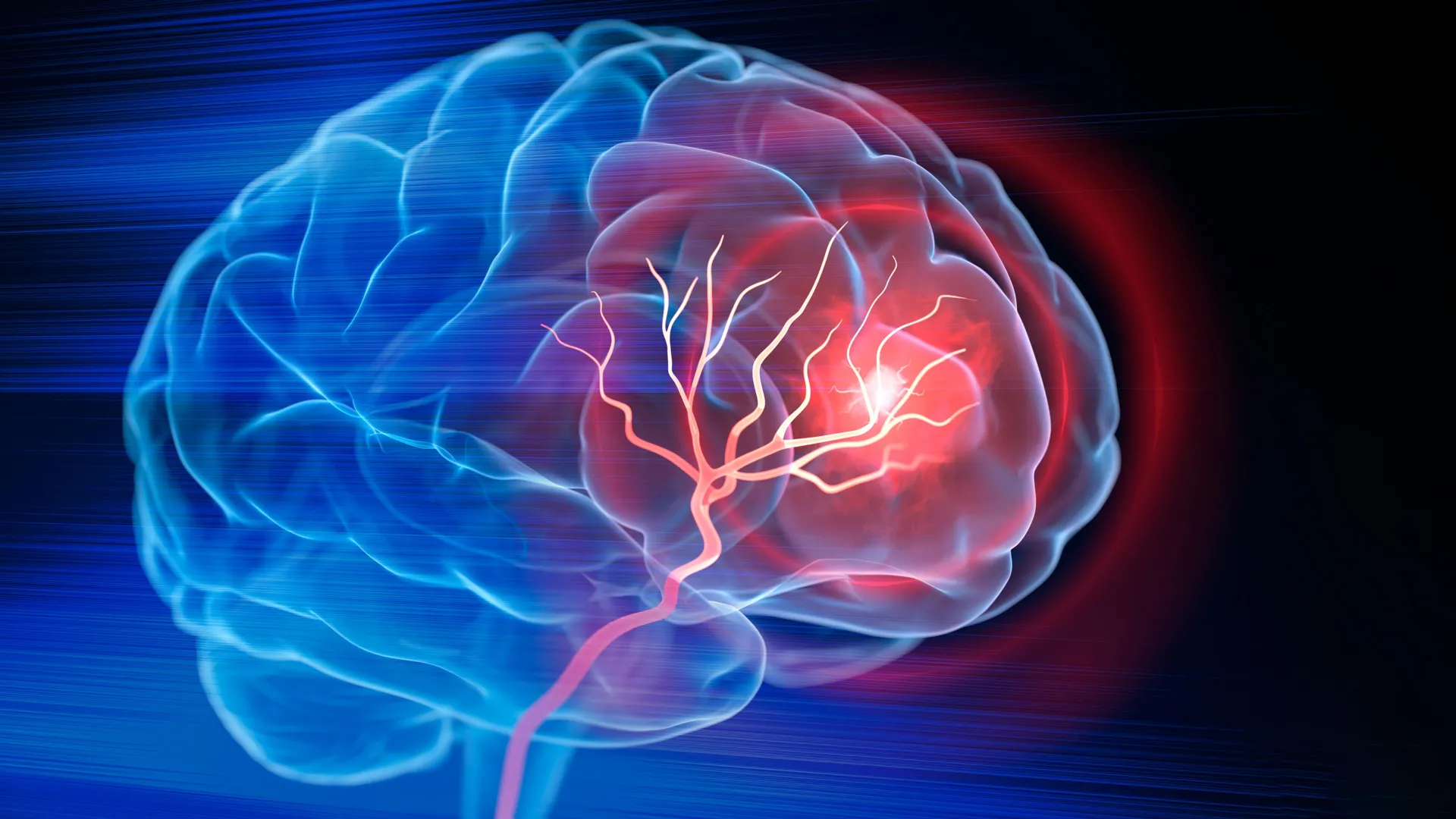
Biomedical engineers at Duke University have developed an injectable biomaterial that could support brain repair after an ischemic stroke. In mouse studies, the treatment transformed cavities left by dead brain tissue into a supportive environment for healing and regeneration.
The injectable brain-repair scaffold mobilized the animals’ own immune cells, encouraged the growth of new blood vessels, supported neural tissue changes, and improved motor function.
The findings highlight the potential of cell-based biomaterials and extracellular vesicle technology for repairing brain damage after stroke.
Why repairing brain tissue after ischemic stroke is difficult
Millions of people experience an ischemic stroke each year. This type of stroke occurs when a blood clot blocks blood flow to part of the brain.
Emergency treatments, including clot-dissolving drugs and procedures that physically remove the blockage, can restore circulation and preserve brain tissue that remains viable. However, restoring blood flow cannot replace brain cells that have already died.
A severe ischemic stroke can destroy a substantial amount of brain tissue, leaving a cavity where healthy tissue once existed. After the clot is removed, recovery typically depends on rehabilitation. Although rehabilitation helps surviving brain circuits adapt, it does not directly rebuild the damaged region.
“Once brain tissue is lost, restoring blood flow is no longer sufficient,” said Tatiana Segura, Robert Pronsey Distinguished Professor of Biomedical Engineering at Duke University. “Our goal is to engineer the injured space so that immune, vascular, and neural repair processes can begin working together.”
Creating an injectable scaffold for brain repair
Segura and her team set out to create a healing environment inside the stroke cavity that could support several repair processes at the same time.
The researchers used MAPS, or microporous annealed particle scaffolds. These scaffolds are made from individual hydrogel microparticles that assemble into a porous structure. The open spaces provide a framework that cells can enter and use as they remodel the damaged area.
Building on earlier work with this biomaterial, the team investigated whether the body’s immune system could be guided to improve tissue repair after stroke.
The researchers focused on astrocytes, star-shaped brain cells that support normal brain function and respond rapidly to injury.
Astrocytes communicate with nearby cells by releasing extracellular vesicles, or EVs. These microscopic packages contain proteins, lipids, and genetic material that can influence the activity of other cells.
Keeping therapeutic repair signals at the injury site
The researchers collected extracellular vesicles from astrocytes grown in the laboratory. They then tested the vesicles with signaling molecules intended to attract immune cells, support blood vessel formation, and improve tissue function.
Instead of injecting the EVs directly into the damaged brain, the scientists chemically attached them to the surface of the hydrogel microparticles. This approach kept the therapeutic signals concentrated inside the scaffold, increasing the likelihood that incoming cells would encounter them.
“We’re not just injecting substances into the brain,” Segura said. “We are designing a local environment where we can coordinate parts of the body’s repair response.”
One combination of signals produced especially promising results. Interleukin-4, or IL-4, and C1q were particularly effective at recruiting potentially beneficial immune cells to the damaged area.
These immune cells included macrophages as well as a surprisingly persistent population of neutrophils.
Neutrophils may support healing after stroke
Neutrophils are commonly associated with inflammation and tissue damage during the early stages of stroke. However, the new findings suggest that their role may change over time.
During later stages of recovery, neutrophils may help support tissue repair when they are exposed to the right signals and material environment.
To test the importance of these cells, the researchers reduced an immune cell population containing many neutrophils. This significantly decreased the formation of new blood vessels, known as angiogenesis, and reduced remodeling of the scaffold.
The results indicate that these immune cells play an important role in the healing response triggered by the biomaterial.
“These results change the way we think about neutrophils after stroke,” said Shangjing Xin, lead scientist on the study and a postdoctoral fellow at the Segura Institute. “Their role appears to depend on when they arrive, where they are located, and the signals they receive from their surroundings. Our study shows the potential of engineering strategies to recruit and maintain these cells at the right time.”
New blood vessels and neural connections formed
As immune cells entered the treated region, new blood vessels formed throughout the stroke cavity.
The researchers also observed more axonal fibers inside and around the injured area. Axons are specialized structures that allow nerve cells to transmit electrical signals.
These biological changes were accompanied by improvements in movement.
Mice treated with the optimized scaffold performed better on a grid-walking test that measured errors in forelimb placement. After eight weeks, their performance was no longer statistically different from that of healthy control mice. The improvement continued for the rest of the study.
The biomaterial scaffold was essential
The researchers also tested whether extracellular vesicles could produce similar effects without the MAPS biomaterial scaffold.
They could not.
EVs delivered without the MAPS scaffold failed to produce comparable improvements in blood vessel repair. This finding suggests that the biomaterial does more than transport therapeutic signals into the brain.
The scaffold’s porous structure, combined with its ability to keep extracellular vesicle signals concentrated at the injury site, appears to be an important part of the repair response.
Injectable stroke treatment remains in the early stages
Despite the promising findings, this injectable brain-repair approach remains in the preclinical stage.
So far, researchers have tested the treatment in mouse models by injecting the biomaterial directly into damaged areas of the brain.
Additional research is needed to evaluate safety, clarify how different immune cell populations affect recovery, and determine whether the treatment works in larger animal models that more closely resemble human stroke.
The researchers are also working to obtain extracellular vesicles from primary rat astrocytes.
As a next step, Segura’s laboratory is studying EVs produced by astrocytes derived from human induced pluripotent stem cells. These cells could give researchers greater control over the signals carried by the EVs while offering a more scalable and clinically relevant source.
“Ecosystems won’t recover just by limiting the initial damage,” Segura said. “We need to create conditions for life to return. That’s how we think about the stroke cavity. This material is not intended to recreate the brain itself, but to create an environment where the body’s own cells can enter, communicate, and participate in the remodeling of vascularized tissue.”
Source: www.sciencedaily.com