


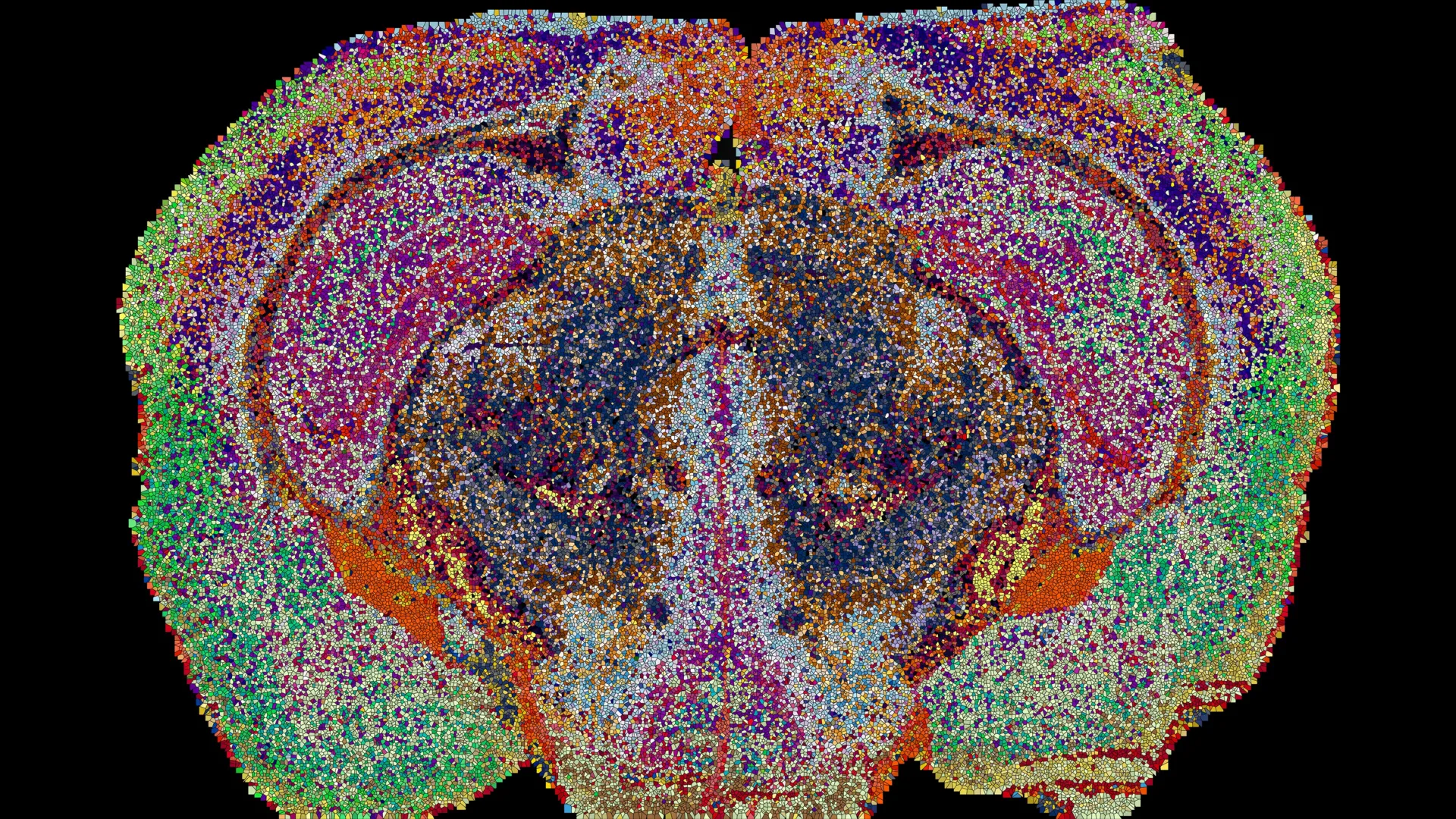
Scientists Map 539 Chemical Zones in the Mouse Brain Using Lipids
Scientists have spent decades studying the brain’s cells, connections, genes, and proteins. Another essential part of brain biology—lipids—has received far less attention. These fatty molecules form the membranes surrounding neurons, contribute to the myelin insulation that protects nerve fibers, and help transmit information between brain cells.
Lipids are fundamental to brain function, but scientists have struggled to determine where different types are located. Traditional imaging techniques cannot easily distinguish individual lipid molecules, leaving major gaps in scientists’ understanding of brain chemistry.
“Scientists have only a vague picture of how lipids differ in one part of the brain from another,” says Professor Giovanni D’Angelo, Chair of the Christian Gerhard Jebsen Foundation for Nutrition and Metabolism at EPFL. “This gap is important because changes in brain lipids are increasingly linked to conditions ranging from depression to Alzheimer’s disease.”
EPFL researchers have now taken a major step toward closing that gap. A team led by D’Angelo, Luca Fusar Bassini, and Gioele La Manno created a detailed map showing how lipids are distributed throughout the mouse brain. The findings were published in Nature.
The new lipid brain atlas revealed that these molecules are arranged in a highly organized pattern. Some patterns match familiar anatomical structures, while others reveal chemical boundaries overlooked by previous approaches. The patterns were so distinctive that researchers could determine where brain tissue samples came from simply by examining their lipid composition.
Scientists identify 539 distinct chemical zones in the brain
To build the atlas, the researchers used mass spectrometry imaging, a technique that identifies lipid molecules across thin sections of brain tissue. A laser scans each slice one location at a time, producing detailed information about which lipids are present and where they are located.
The research team examined 172 different lipids in 109 brain slices taken from 11 mice. This generated approximately 7 million measurements, which scientists integrated into a comprehensive three-dimensional reconstruction using machine-learning tools developed specifically for the project.
The resulting atlas revealed 539 chemically distinct regions, which the researchers named “lipizones.” Each lipizone has a characteristic combination of lipids that creates a recognizable chemical signature.
Many of these regions correspond to established brain structures and cell types. Others reveal unexpected relationships between groups of neuronal cell bodies and distant regions connected by their fibers. These associations can be difficult to detect using maps based only on gene activity.
“Lipids actually act like zip codes, giving distant but related parts of the brain the same chemical address,” D’Angelo says.
Hidden lipid patterns challenge conventional brain maps
The atlas also revealed surprising differences within brain regions that scientists previously considered relatively homogeneous.
One prominent example is white matter, the tissue containing nerve fibers that connect different brain regions. Rather than having a roughly consistent chemical composition, white matter contains numerous distinct lipid zones.
“This shows that the chemistry of the cells that make myelin is much more diverse than genetics alone would suggest,” says Luca Fusar-Bassini.
The researchers also detected previously unrecognized chemical boundaries in the choroid plexus, a structure involved in producing cerebrospinal fluid, and in the tissue lining the brain’s internal cavities.
Taken together, the findings indicate that lipid mapping can reveal important features of brain tissue that are not evident from anatomy or genetic information alone.
Pregnancy causes widespread changes in brain lipids
In addition to identifying the brain’s chemical structure, the atlas provided new insight into how that structure changes during pregnancy.
When researchers examined pregnant mice, they found significant changes in lipid composition across numerous brain regions. Overall, these changes were more pronounced than the typical lipid differences observed between male and female mice.
Particularly extensive changes occurred in the cortex, which forms the brain’s outer layer. Levels of galactosyl ceramide, a key component of myelin, also increased sharply throughout most of the brain’s white matter.
These findings suggest that pregnancy involves significant changes in the brain’s chemical composition. Such changes may help neural connections adapt to the body’s changing demands.
“Taken together, these changes suggest that the brain is rewiring itself to meet the demands of pregnancy,” Gioele La Manno says. “The atlas reveals that the brain constantly adjusts lipids to match the body’s changing needs.”
A new tool for studying brain aging and disease
The Lipid Brain Atlas is available for free online, allowing scientists around the world to investigate this previously difficult-to-study aspect of brain biology.
Combining lipid information with existing genetic and cellular maps could help researchers develop a more complete understanding of how the brain is organized and how its chemistry changes over time.
The atlas also provides a detailed reference for healthy mouse brain tissue. Scientists can use it to investigate how disruptions in lipid distribution contribute to brain development, aging, and diseases such as Alzheimer’s disease and depression.
Although a mouse brain map cannot be directly applied to humans, the method used to create it could help researchers uncover comparable patterns in the human brain.
“What is transmitted to humans is not a map, but a method, and the fact that we know that healthy brains have very organized lipid maps,” says Luca Fusar-Bassini.
Source: www.sciencedaily.com