


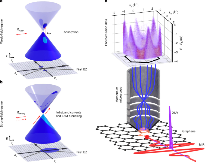
The experiment combines high-repetition-rate extreme-ultraviolet (XUV) pulses with intense, phase-stable mid-infrared (MIR) fields to investigate field-driven electron dynamics in monolayer graphene. A time-of-flight momentum microscope records the emitted electrons with broad momentum coverage, while electro-optic sampling reconstructs the MIR waveform. The graphene samples are epitaxial monolayers grown on silicon carbide and are measured under ultrahigh vacuum at room temperature.
Experimental setup
The subcycle momentum microscopy setup is based on two ytterbium-doped potassium gadolinium tungstate (Yb:KGW) amplifiers (CARBIDE, Light Conversion Inc.). Each amplifier produces near-infrared pulses with a duration of 250 fs, a centre wavelength of 1,030 nm and a pulse energy of 1.5 mJ at a repetition rate of 50 kHz.
In the probe arm, the second harmonic of a four-stage noncollinear optical-parametric amplifier (NOPA; ORPHEUS-OPCPA, Light Conversion Inc.)48 produces carrier-envelope-phase (CEP)-stabilized pulses centred at 400 nm, with a duration of 20 fs. These pulses are focused into a high-pressure argon gas jet to generate high-order harmonics in a vacuum beamline49. Two multilayer mirrors spectrally select the seventh harmonic, which has a photon energy of 21.7 eV. The mirrors have a bandwidth of 0.9 nm around 57 nm, supporting a Fourier-limited pulse duration of less than 10 fs. The XUV pulses enter the experimental ultrahigh-vacuum chamber and momentum microscope32 through a 200-nm-thick aluminium foil, which also removes the ultraviolet driving pulses.
Because a time-of-flight photoemission momentum microscope detects only about one electron per laser shot, a high repetition rate is essential for obtaining good statistics within a reasonable measurement time. This requirement makes the generation of atomically strong, few-cycle, phase-stable MIR pump pulses a significant challenge for a tabletop system. The phase-stable MIR pulses used here have, to the best of the authors’ knowledge, record-high average powers of up to 1 W and electrical peak field strengths exceeding 200 MV cm−1.
The MIR field transients are generated by difference-frequency generation in a gallium selenide crystal using the outputs of two parallel (N)OPAs (ORPHEUS-MIR, Light Conversion Inc.)50. The pulses have durations as short as 50 fs and can be tuned from 4 µm to 15 µm. A small portion of the fundamental laser output is directed to a home-built single-stage NOPA, optimized to generate 10-fs pulses centred around 825 nm. These pulses are used for electro-optic sampling of the MIR waveforms. Extended Data Fig. 1c shows a typical MIR transient measured by electro-optic sampling. A pair of wire-grid polarizers controls the MIR polarization direction and power before the pulses are coupled into the ultrahigh-vacuum chamber through a diamond window.
The intense, s-polarized MIR pump and the p-polarized XUV probe are spatially overlapped in the vacuum chamber using a custom-built off-axis parabolic mirror with a small aperture for XUV transmission. They are focused onto monolayer graphene at an angle of incidence of 70°. A piezo-controlled delay stage in the excitation beam path sets the delay between the MIR and XUV pulses with a temporal precision of 230 as.
The emitted photoelectrons are measured with a time-of-flight momentum microscope equipped with a lens system from GST mbH and a HEX40 detector from RoentDek51. Multihit detection maintains signal linearity at repetition rates of up to 20 kHz. The 21.7-eV probe pulses, combined with the microscope’s large acceptance angle, provide access to momentum regions extending to ±2.14 Å−1. This is substantially broader than the ±0.16 Å−1 range available in earlier subcycle angle-resolved photoemission spectroscopy experiments8,20.
The energy resolution is limited by the broad probe-pulse spectrum. The XUV bandwidth can be tuned, giving an energy-resolution limit of 180 meV. The momentum resolution is better than 0.01 Å−1. The custom-designed ultrahigh-vacuum chamber is shielded by a μ-metal liner and has a base pressure of 2 × 10⁻¹⁰ mbar. Measurements were performed at room temperature.
Driving field-dependent dynamics in the monolayer requires the electric field to be parallel to the graphene plane. The MIR pulse is therefore s-polarized to maximize this field component. The XUV polarization is perpendicular to the MIR polarization, preventing field crosstalk that could produce coherent artefacts in the observed dynamics.
The sample was also rotated so that MIR-induced momentum streaking in vacuum was minimized at one of the K points. This alignment places one K point at k∥ = 0, where k∥ is the momentum component parallel to the MIR electric field. It also reduces matrix-element-induced asymmetries along k∥ = 0 in the measured energy and momentum range.
An asymmetry along the kx direction may remain, but it is relatively small at the observed energies. The dark corridor begins to appear only at comparatively high excitation energies, which do not occur along the kx direction in this experiment. This may also account for the slightly greater broadening seen in simulations that neglect the matrix element.
Graphene samples
An n-doped 6H-SiC(0001) wafer was used as the substrate for growing an epitaxial monolayer of graphene with 0° rotational alignment relative to the substrate lattice, referred to as 0°-EMLG. The wafer was first degassed in ultrahigh vacuum at 880 °C for 30 min. Its temperature was then raised to 1,050 °C and held for 30 min under an external silicon flux, producing a silicon-rich (√3 × √3)R30° surface reconstruction.
The sample was subsequently annealed at 880 °C for a further 30 min, again under a silicon flux, resulting in a silicon-rich (3 × 3) surface reconstruction. This procedure removes the oxide layer from the SiC surface and produces an atomically clean surface.
To form 0°-EMLG, the sample was annealed at 1,400 °C for 30 min in a borazine (B3N3H6) atmosphere with a partial pressure of 1.5 × 10⁻⁶ mbar. The borazine molecules act as surfactants on the heated SiC surface. They suppress the formation of the conventional 30°-rotated graphene layer and instead promote a homogeneous graphene layer with the unconventional 0° orientation relative to the substrate lattice. Further details of the 0°-EMLG preparation are given in refs.38,52,53.
After growth, the surface morphology and crystalline structure were characterized using low-energy electron microscopy and low-energy electron diffraction. The SiC surface had an average terrace width of approximately 160 nm and was fully covered by 0°-EMLG.
The sample was then briefly exposed to air, transported in static vacuum at 0.1 mbar to the experimental chambers, and degassed at 350 °C for 3 h before measurement. Photoemission measurements showed that the 0°-EMLG was n-doped, with the Fermi level approximately 200 meV above the Dirac point.
Electric-field waveform reconstruction and streaking compensation
The acceleration of photoemitted electrons in vacuum enables in situ reconstruction of electric-field waveforms20. In addition to acceleration inside the sample, the s-polarized field accelerates electrons after they have left the material. This produces a momentum change of the entire band structure parallel to the sample surface, Δk∥, which follows the vector potential of the field. The electric-field waveform can therefore be calculated from this momentum change.
Source: www.nature.com