


Scientists Identify Drug-Targetable Receptor That May Reduce Brain Inflammation
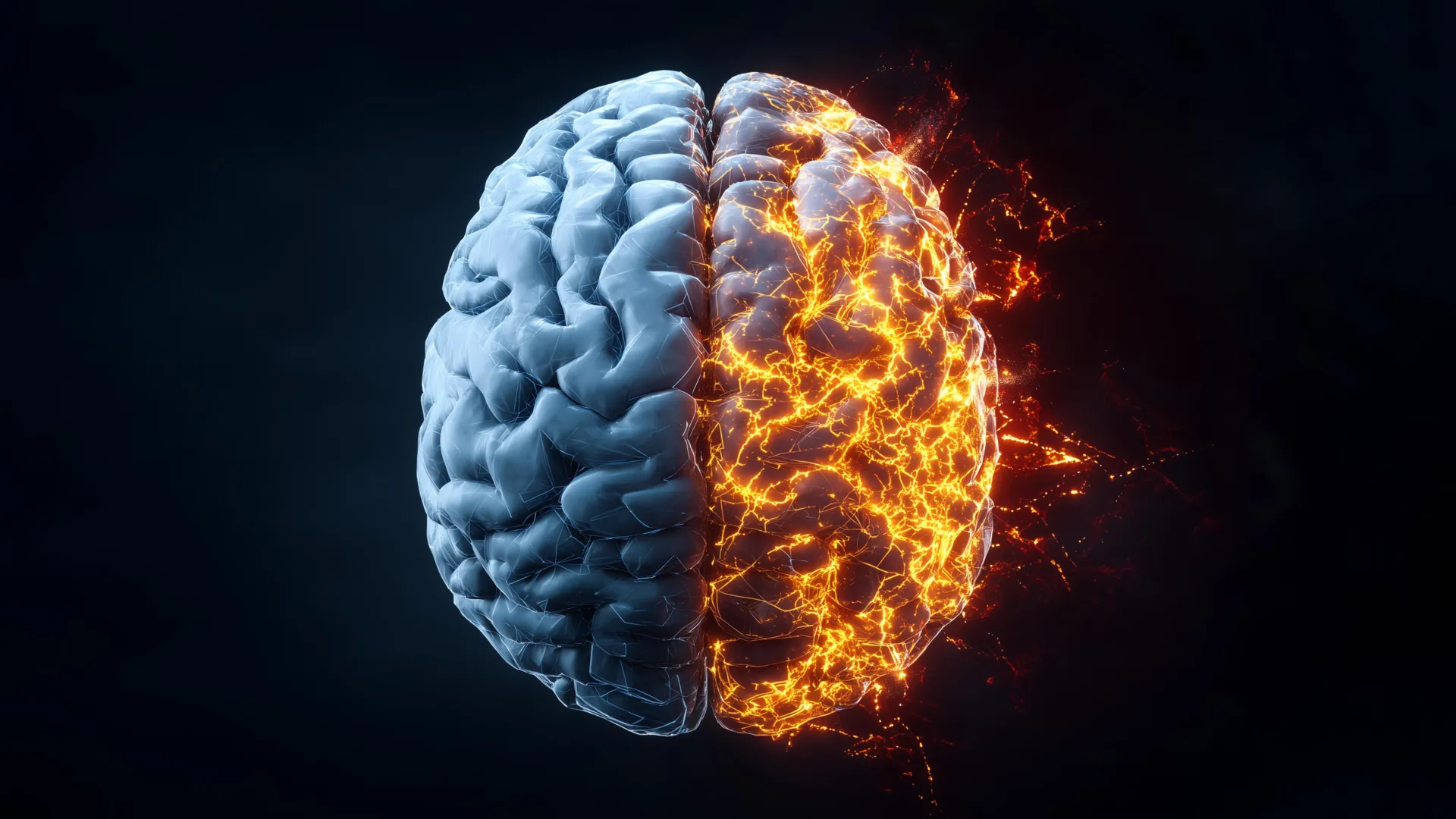
Scientists may have discovered a new way to reduce harmful inflammation in the brain by targeting P2X7 receptors, which can already be blocked by existing drugs.
Researchers led by Professor Nicholas Burns at the University of Birmingham identified P2X7 receptors as a key factor in neuroinflammation. When the researchers blocked this receptor, inflammation in human brain tissue was significantly reduced.
The discovery could eventually have implications for neurological and psychiatric conditions in which inflammation is thought to play a role. These include traumatic brain injury (TBI), Alzheimer’s disease, Parkinson’s disease, depression and psychosis.
P2X7 receptor linked to brain inflammation
To investigate the receptor, the researchers used living cultures of human brain cells and slices of brain tissue collected during neurosurgery. They focused on P2X7, a receptor that helps trigger inflammatory signaling.
The research team found that P2X7 receptors promote the release of cytokines, proteins that help control inflammatory responses. When the researchers used a specific P2X7 antagonist to block the receptor, the inflammatory response in human brain tissue was significantly reduced.
Professor Nicholas Burns, from the School of Medicine and Health at the University of Birmingham and corresponding author of the paper, said:
“This exciting discovery marks a major step towards repurposing existing treatments to fight neuroinflammation at its roots. The identification of this receptor could have far-reaching implications for some of the most debilitating and widespread brain disorders, such as Alzheimer’s disease, Parkinson’s disease and multiple sclerosis, or inflammation-related psychiatric diseases, such as schizophrenia and depression.”
Researchers study the brain’s immune cells
Much of the research focused on microglia, immune cells that help coordinate the brain’s response to injury and inflammation.
To study how these cells respond to inflammatory signals, the researchers developed a method to convert certain white blood cells into microglia-like cells. This process reflects cellular changes recently identified as occurring naturally in the brain during human aging.
The research team began with human peripheral monocytes taken from blood samples and converted them into microglia-like cells. These cells provided a way to examine how human microglia respond to inflammatory signals.
When the researchers applied a P2X7 receptor antagonist, they were able to block the signals released by microglia when the cells were damaged and died.
Professor Burns said:
“Studying human microglia has long been a major challenge. When human microglia are removed from their native brain environment, they rapidly lose their characteristics, presumably due to the absence of important regulatory signals. Our approach involves the use of monocyte-derived microglia, which provides a powerful, scalable and virtually unlimited platform to study human microglial biology with unprecedented precision.”
Findings replicated in human brain tissue
After identifying the response in microglia-like cells grown in the laboratory, the researchers tested whether the same findings could be replicated in human brain tissue obtained during neurosurgery.
The successful results in human tissue strengthen the case for further investigation and may eventually lead to clinical trials in people with neurodegenerative diseases or traumatic brain injury.
Professor Burns said:
“Identifying the response of human monocyte-derived microglia has led us to translate these findings after neurosurgery to the human brain. This successful translation means that the next step in this research is the development of clinical trials in patients with neurodegenerative diseases and traumatic brain injury, where there are no effective pharmacological treatments to reduce neuroinflammation and the damage that is occurring.”
Source: www.sciencedaily.com