


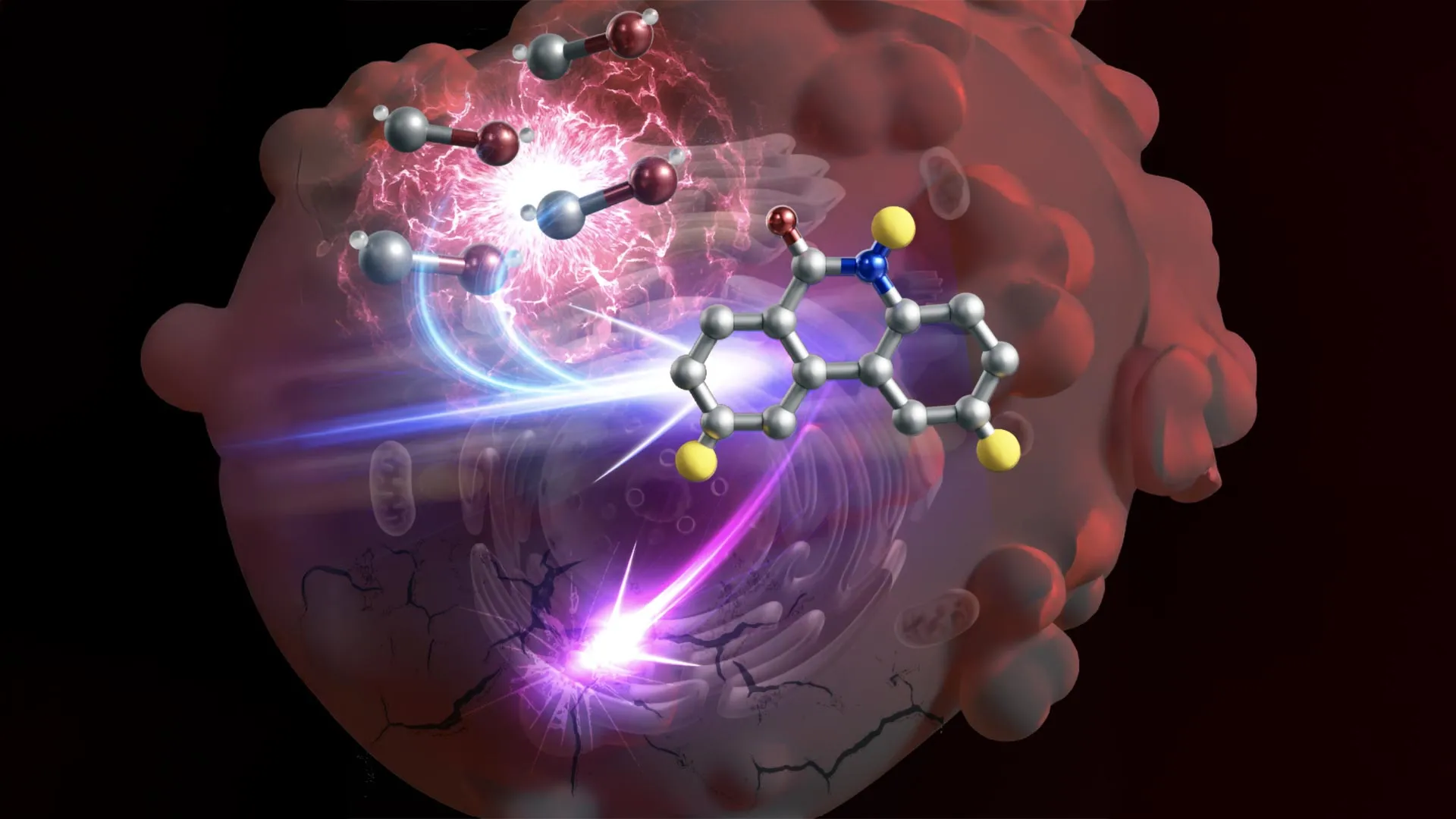
Japanese researchers have developed an experimental strategy that uses carbon monoxide produced by cancer cells to manufacture anti-cancer compounds inside those cells. Led by Katsunori Tanaka of the RIKEN Pioneering Research Center (PRI), the team showed in laboratory experiments that a specially designed compound could trigger drug production in several cancer cell types without generating the same toxic substances in non-cancerous cells.
Turning a cancer byproduct against tumors
One of the central challenges in cancer treatment is destroying malignant cells without harming healthy tissue. Conventional chemotherapy can cause severe side effects because its toxic effects are not necessarily confined to tumors. Tanaka and his colleagues are investigating more selective approaches that could make treatment safer.
Their long-term goal is to replace the direct delivery of toxic drugs with relatively harmless compounds that become active only after entering cancer cells. These compounds would use substances already present within tumors to produce therapeutic agents where they are needed.
Carbon monoxide is a potential target for this approach. The gas is best known as a toxic pollutant produced by sources such as gasoline engines, but many types of cancer cells also produce it naturally. Because tumors can generate unusually large amounts of carbon monoxide, the gas could serve as a biological marker for identifying cancer cells.
Scientists at RIKEN’s Biofunctional Synthetic Chemistry Laboratory investigated whether this difference could be exploited. Their strategy involved delivering metal catalysts into cells and using the excess carbon monoxide in cancer cells to trigger chemical reactions that produce toxic compounds. In principle, the reaction should occur mainly in cells containing sufficient carbon monoxide, while leaving normal cells unharmed.
Protecting the catalyst inside cells
The researchers used a group of compounds known as palladacycles. These metal-containing compounds react with carbon monoxide to produce organic molecules called phenanthridinones. Some phenanthridinone derivatives are toxic to cells and have already been incorporated into anti-cancer drugs.
However, the approach faced a significant obstacle. When a palladacycle is introduced directly into cells, substances such as glutathione can rapidly deactivate its central palladium catalyst. Without an active catalyst, the reaction cannot produce the intended drug.
Tanaka’s team built on earlier work involving metal catalysts in biological systems. They encapsulated the palladacycle in albumin, a protein commonly found in blood. The protein carrier allows the complex to travel through the bloodstream while helping protect the palladium catalyst from substances that could inactivate it after entering a cell.
Drug production in cultured cancer cells
The researchers first tested several palladacycles to identify the compound that produced the greatest amount of phenanthridinone.
They then exposed cultured human liver, cervical, breast and lung cancer cells to albumin–palladacycle complexes for 48 hours. Non-cancerous cell lines were also tested to determine whether the same reactions occurred in healthy cells.
After 48 hours, the researchers detected phenanthridinone in the cancer cell lines but not in the normal cells. The result indicated that the albumin carrier had helped protect the catalyst from glutathione and that, under the conditions tested, the chemical reaction occurred selectively in cancer cells.
The team further refined the system so that the newly formed compounds could separate from the carrier and move freely within cells. Additional modifications allowed the reaction to produce particularly toxic phenanthridinone derivatives that are already used in anti-cancer drugs.
When the modified system was introduced into cultured liver cancer cells, the researchers observed anti-cancer compounds forming inside the cells. The cells subsequently showed signs of damage caused by the toxic drug, providing evidence that the method can generate cancer-fighting substances directly at the intended target.
Why the drug did not form in blood
A key safety concern was whether carbon monoxide in the bloodstream could trigger the reaction before the compound reached cancer cells. If that happened, the resulting drug could damage blood cells instead of the tumor.
Laboratory tests showed that toxic drugs were not produced in blood. The researchers believe this is because most carbon monoxide in the bloodstream is bound to hemoglobin, the protein responsible for transporting oxygen. In this bound form, the gas does not appear to be available for the reactions required to manufacture the drug.
The findings suggest that the system may be able to travel through the blood without producing harmful substances prematurely. However, further research is needed to establish whether it is safe in living organisms.
“In our therapeutic strategy, molecules that are naturally present or produced in the body, including disease-related metabolites, are used as endogenous chemical raw materials to synthesize therapeutic molecules directly at the site of disease,” Tanaka says.
The research, reported in Nature Communications, remains at an early stage. The team plans to move beyond cultured cells and test the approach in animal models. If those experiments are successful, the technology could contribute to a class of cancer treatments that use tumor chemistry to produce drugs directly at the disease site, potentially limiting damage to healthy tissue.
Source: www.sciencedaily.com