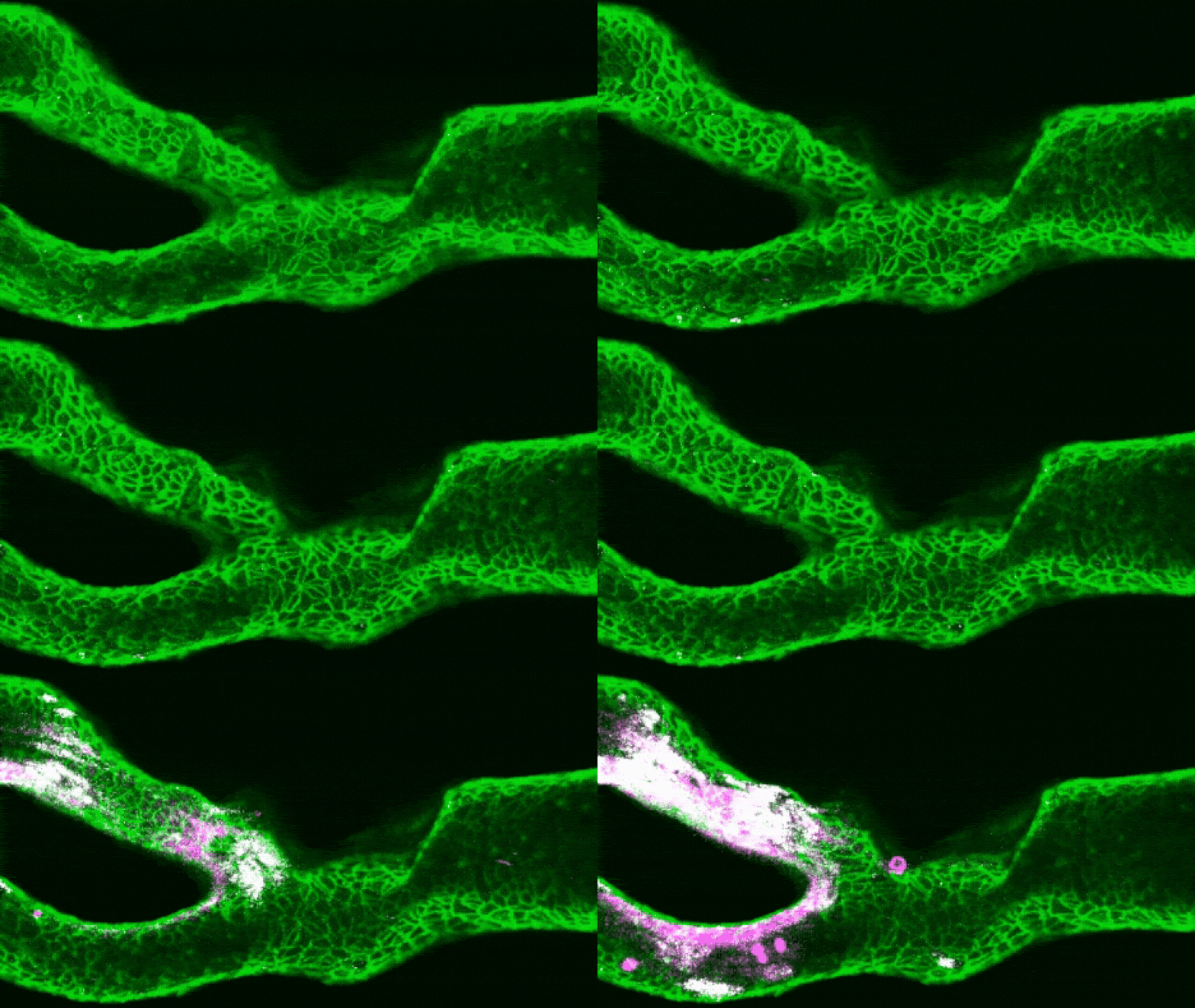
Scientists have developed miniature, patient-specific models of the carotid artery that could help doctors predict stroke risk and choose more effective treatments for blood clots, according to a new study.
Published in Cell Biomaterials in July, the research describes an “artery-on-a-chip” system designed to recreate how blood flows through an individual patient’s carotid artery. These major blood vessels carry oxygen-rich blood to the brain, face and neck.
The technology could give clinicians a closer look at how a patient’s blood clots form, grow and break apart. It may also help identify which medications are most likely to prevent a dangerous blockage.
Although the artery-on-a-chip technology is still a proof of concept, it could eventually support more personalized stroke prevention and treatment.
from the latest videolive science
How artery-on-a-chip models could improve stroke care
“We believe this approach is most relevant for difficult clinical cases,” said study lead author Charles Chao, a PhD student at the University of Sydney. The models could be especially useful for patients whose condition returns despite treatment or for cases in which doctors must choose between several treatment strategies, Zhao told Live Science in an email.
Ischemic stroke is among the leading causes of death worldwide. It occurs when a blood clot blocks an artery supplying the brain, depriving brain cells of oxygen. High blood pressure, obesity and high cholesterol can increase the risk. Atherosclerosis — the buildup of fatty plaques inside artery walls — contributes to approximately one in five ischemic strokes.
Why blood clot behavior matters
When the inner lining of an artery is damaged, proteins such as collagen beneath the cells become exposed to the bloodstream. A blood protein called von Willebrand factor, or VWF, captures platelets as they move through the vessel. The platelets then attach to one another and attract additional platelets, creating a blood clot.
However, the size of an arterial blockage is only part of the stroke risk. The way a clot behaves — whether it remains stable or breaks into smaller fragments — can determine what happens next.
Get the world’s most fascinating discoveries delivered straight to your inbox.
Some clots remain attached to the artery wall and only partially restrict blood flow. Others can shed small pieces that travel toward the brain. If one of these fragments blocks a smaller blood vessel, it can cause an ischemic stroke.
“Clinically, we’re very good at imaging how narrow an artery is, but narrowing alone doesn’t tell us exactly how a blood clot will behave,” Zhao said.
Creating a personalized carotid artery model
The researchers used 3D printing and patient CT scans to recreate the precise shape of each participant’s carotid artery, including areas narrowed by atherosclerotic plaque. They printed a plastic replica, coated its interior with collagen and added the cells that normally line the artery.
Blood was then pumped through the model at speeds and pressures designed to mimic real blood flow. This allowed the researchers to observe how clots developed under conditions that closely matched each patient’s vascular anatomy.
Study co-author Zihao Wang holds an assembled 3D-printed vascular device.
(Image credit: University of Sydney/Fiona Woolf)
To simulate an injury, the team used a small laser to expose the collagen beneath the artery’s lining. Human blood was then circulated through the model, allowing the formation of a clot to be monitored under a microscope, Zhao explained.
The researchers built artery-on-a-chip models for six patients with different types of arterial damage. They combined observations from the models with calculations showing how changes in blood flow affected clot formation and movement.
Patient-specific models reveal differences in clot stability
The results showed that medical images alone may not capture important differences in how blood clots develop. Even patients with apparently similar arterial disease can have very different clotting behavior.
“We found that two arteries with seemingly similar diseases can behave very differently because their three-dimensional shapes create different blood-flow patterns,” Zhao said. These localized flow conditions can influence whether a clot grows in a stable manner or becomes vulnerable to breaking apart.
The findings could eventually help doctors select treatments based on how a specific patient’s clots form. Some medications reduce platelet clumping, while others prevent platelets from attaching to artery walls or block VWF, a key protein involved in clot formation.
Because the most effective medication may depend on the biological and mechanical behavior of an individual’s clot, patient-specific artery-on-a-chip models could complement traditional CT and other medical imaging techniques, Zhao said.
Next steps for artery-on-a-chip stroke research
The researchers plan to expand the study and investigate whether artery-on-a-chip testing could become an additional diagnostic step between initial medical imaging and treatment selection.
More research involving larger numbers of patients will be needed before the technology can be evaluated for routine clinical use. For now, the study demonstrates how combining 3D printing, patient imaging and real blood flow could provide new insight into stroke risk.
This article is for informational purposes only and does not provide medical advice.
What do you know about the hardest-working muscles in your body? Find out with our Heart quiz!