


More than 10,000 people with chronic liver disease are waiting for a liver transplant in the United States, yet the supply of donated organs remains far below demand. Some patients with liver failure are also too sick to safely undergo transplant surgery, creating an urgent need for alternative treatments.
MIT engineers are developing a potential solution: an injectable “mini-liver” designed to restore some of the vital functions normally performed by a damaged liver.
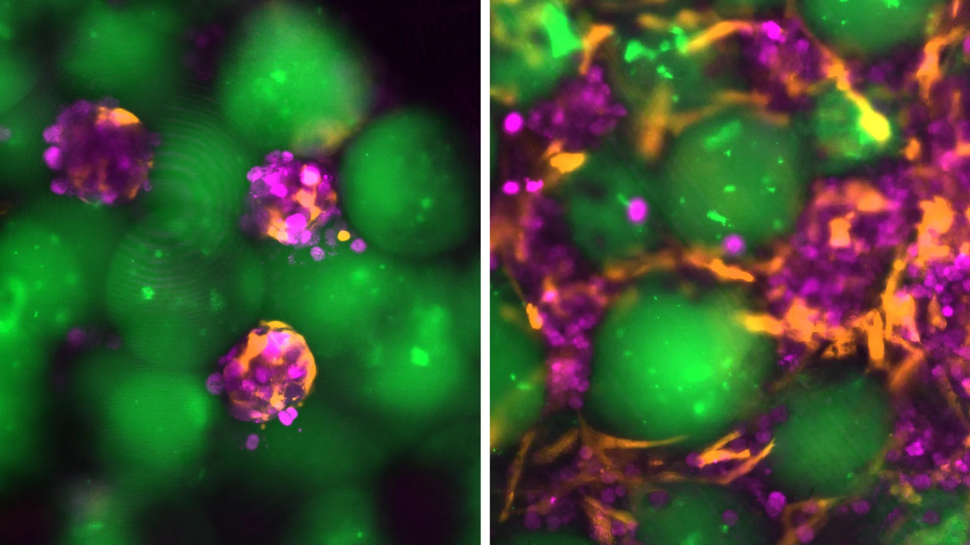
In a new study involving mice, researchers found that injected liver cells remained alive and functional for at least two months. During the study, the cells continued producing many of the enzymes and proteins typically made by healthy liver tissue.
“We think of these as satellite livers. If we can deliver these cells into the body while leaving the diseased organ in place, it could provide a booster function,” says Sangeeta Bhatia, the John and Dorothy Wilson Professor of Health Sciences and Technology and of Electrical Engineering and Computer Science at MIT. Bhatia is also a member of MIT’s Koch Institute for Integrative Cancer Research and the Institute for Biomedical Engineering and Science (IMES).
Bhatia is the study’s senior author, and MIT postdoctoral fellow Vardhman Kumar is the lead author. The findings were published in Cell Biomaterials.
Restoring vital liver function
The liver performs approximately 500 essential functions in the human body. It helps regulate blood clotting, remove bacteria from the bloodstream, process medications, and carry out many other critical tasks. Many of these functions are performed by specialized liver cells called hepatocytes.
For more than a decade, Bhatia’s laboratory has explored ways to restore liver function without requiring patients to undergo surgical liver transplantation. One approach involves placing hepatocytes inside biomaterials such as hydrogels. However, these materials still require surgical implantation.
Injecting liver cells directly into the body could eliminate the need for that procedure. In the new study, the MIT team developed an engineered environment that helps transplanted cells survive and enables doctors to monitor the graft using noninvasive imaging.
Creating an injectable environment for liver cells
The researchers created an injectable mixture containing liver cells and tiny hydrogel microspheres. These microspheres help support the cells and promote connections with nearby blood vessels.
When densely packed, the microspheres behave like a liquid, allowing the mixture to pass through a syringe. After injection, the material recovers its solid structure and provides a stable environment for the transplanted cells.
Hydrogel microspheres have previously been studied for wound healing because they create spaces that allow cells to move through the material and form new tissue. The MIT researchers adapted this technology to help liver cells establish stable tissue grafts after injection.
“What we’ve done is use this technology to create an artificial niche for cell transplantation,” Kumar says. “If we inject cells without these spheres, the cells will not integrate efficiently with the host. These microspheres provide a niche for the hepatocytes, allowing them to remain localized and connect with the host circulation much faster.”
The injectable material also contains fibroblasts, supportive cells that improve liver cell survival and encourage blood vessel growth within the newly formed tissue.
Working with Nicole Henning, an ultrasound research expert at the Koch Institute, the team developed a technique for delivering the mixture with an ultrasound-guided syringe. Ultrasound can also be used to monitor the implant after injection and determine whether it remains stable over time.
Injectable mini-liver may work in different parts of the body
In mouse experiments, the researchers implanted the mini-livers in fatty tissue inside the abdomen. Future versions of the treatment could potentially be placed in other areas, including regions near the spleen or kidneys.
The transplanted liver cells do not necessarily need to be located next to the patient’s liver. As long as the graft has enough space and receives an adequate blood supply, the cells may perform functions similar to those of liver cells inside the organ.
“For the majority of liver diseases, there is no need to place the graft near the liver,” Kumar says.
New blood vessels support mini-liver survival
During the mouse experiments, researchers injected the liver cells and hydrogel microspheres into fatty tissue known as perigonadal adipose tissue. After injection, the cells organized into stable, compact structures.
Over time, blood vessels grew into the graft, providing nutrients and oxygen that helped the transplanted liver cells survive and remain functional.
“The liver cells were able to survive because new blood vessels formed right next to them,” Kumar says. “These vessels delivered nutrients effectively, allowing the cells to function normally and produce the proteins we expect.”
The liver cells survived throughout the eight-week study and continued releasing specialized proteins into the animals’ bloodstream. According to the researchers, this durability suggests that the injectable mini-liver technology could eventually be developed as a long-term treatment for liver disease.
Potential bridge to liver transplantation
Researchers see several possible applications for the technology. For some patients, an injectable liver cell graft could eventually offer an alternative to transplant surgery. For others, it could temporarily support liver function while they wait for a donor organ.
“We believe this technology can not only provide an alternative to surgery, but also serve as a bridge to transplantation, where the graft can provide support until donor organs become available,” Kumar says. “If a patient needs another treatment or additional grafts, the injection technique could make that process much easier than undergoing another surgery.”
Current versions of the treatment would likely require immunosuppressive drugs to prevent the immune system from attacking the transplanted cells. The research team is investigating additional strategies, including the development of “stealth” liver cells that can avoid immune detection. Another potential approach is using hydrogel microspheres to release immunosuppressive medication directly around the graft.
This research was funded by the National Cancer Institute, the National Institutes of Health, the Wellcome Leap HOPE program, a National Science Foundation Graduate Research Fellowship, and a Koch Institute Support (Core) grant from the Howard Hughes Medical Institute.
Source: www.sciencedaily.com