


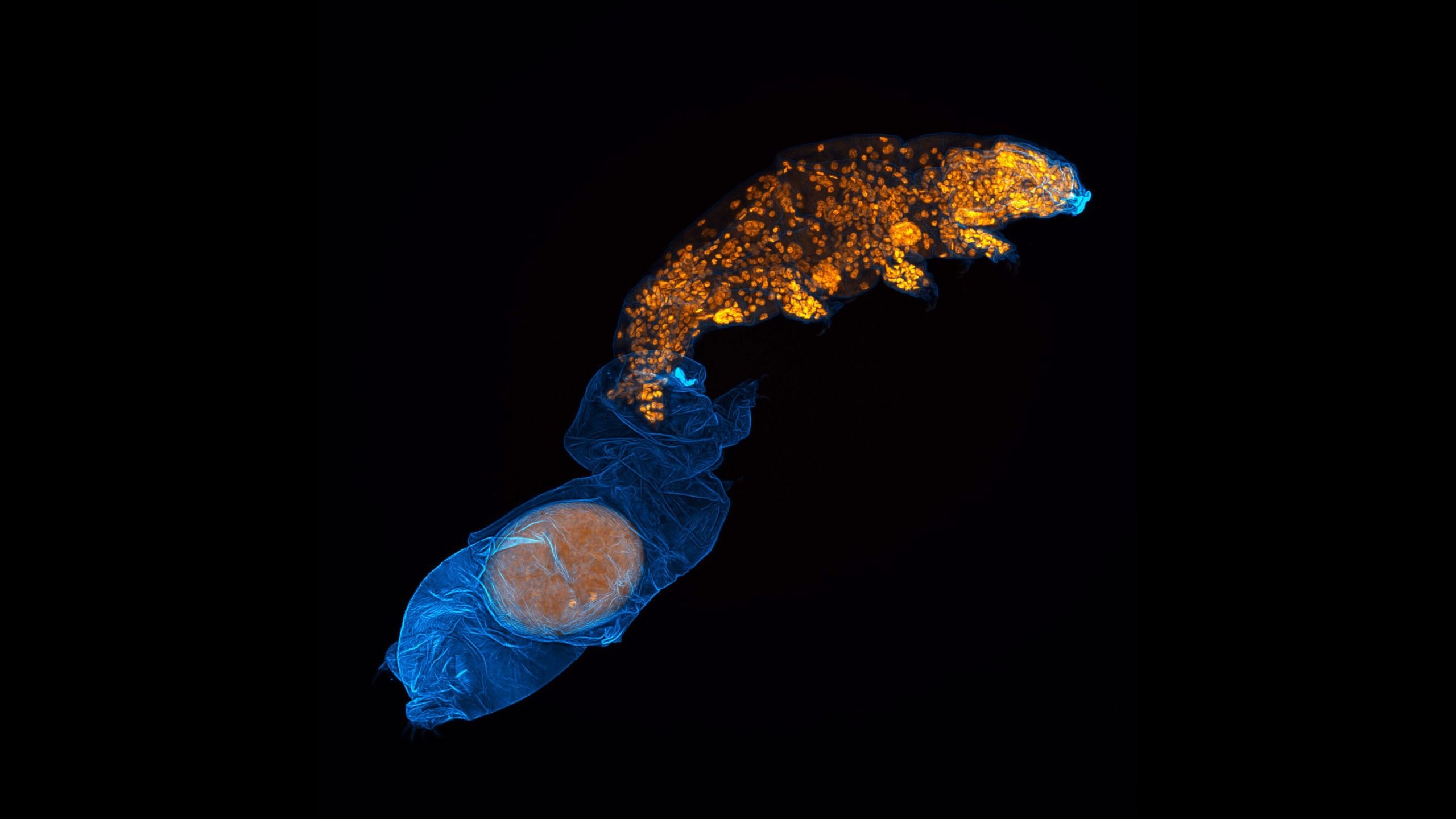
Microscope images by biologist Gonzalo Quiroga-Artigas offer a rare, detailed look at the hidden life of tardigrades. One of his photographs, which placed 12th in Nikon’s 2026 Microscope Photography Contest, shows a Hypsibius exemplaris tardigrade shedding its hard outer layer while laying eggs inside the discarded skin.
A protected nursery inside a molt
Quiroga-Artigas, a postdoctoral researcher at the Montpellier Center for Cell Biology in France, told Live Science that the image emerged during long sessions at a confocal microscope.
“A large part of my research involves imaging, and while spending hours with a confocal microscope, I encountered some incredibly beautiful and unexpected sights,” he said.
“An interesting feature of tardigrade reproduction is that some species lay their eggs inside the molt, or molt layer. The molt layer can therefore provide an additional layer of physical protection to the developing embryo.”
In some species, males may deposit sperm inside the cuticle of a female before she sheds it, according to National Geographic. In other species, a male fertilizes the eggs after they have been left inside the shed cuticle. Some tardigrades do not require a male to reproduce.
“Many species breed” through parthenogenesis, Quiroga-Artigas added. “Parthenogenesis refers to the development of an embryo from an unfertilized egg, also known as a virgin pregnancy. This means that a male is not needed for reproduction and the resulting offspring are genetically very similar to the mother.” The species shown in the photograph belongs to this category.
Molting and spawning can take place over several hours, but the molt itself is relatively quick, making the process difficult to capture on camera.
Finding the right tardigrade
“Once we collected the egg-laying animals, we had to fix, stain and mount the specimens before we could image them,” Quiroga-Artigas said. “We then spent long microscopy sessions looking for the right specimen and the right orientation to obtain high-quality images. Finding specimens that are at exactly the right stage of molting and in an orientation that allows us to clearly visualize the process is largely a matter of patience and luck.”
The animals’ tiny size adds to the challenge. “Tardigrades are very small, so working with tardigrades in the laboratory requires a lot of experience with micromanipulation,” Quiroga-Artigas said. Hypsibius exemplaris, for example, is only about 250 micrometers wide—about twice the width of a human hair.
The photograph was taken with a confocal microscope. The instrument uses a focused laser beam to illuminate one section of a specimen at a time, producing detailed, high-resolution images. Quiroga-Artigas used the same technique to create another tardigrade photograph that was named the best image in the contest.
Beyond still images
Quiroga-Artigas also submitted a video to the Nikon Small World in Motion Competition. It shows a pregnant tardigrade coming into contact with another tardigrade, which then defecates. Quiroga-Artigas said it is unclear whether the contact caused the movement.
He hopes his photographs and videos will show viewers the beauty and complexity of the microscopic world.
“There are tiny animals all around us that we can’t see with the naked eye, but they live, reproduce, grow, and go through complex life cycles just like the animals we’re familiar with,” he said.
“We will continue to image tardigrades in a variety of situations that will be relevant for future research, without necessarily having a specific imaging goal in mind,” he added. “In my experience, some of the best images I’ve taken weren’t necessarily ones I was actively looking for.”
Source: www.livescience.com