


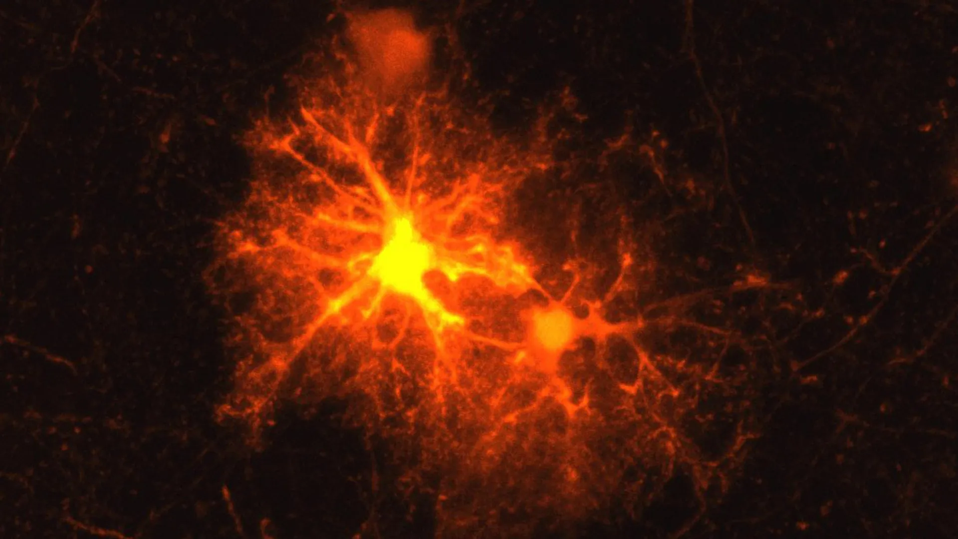
When laboratory mice suffered brain injuries, researchers repeatedly observed the same response: specific groups of cells gathered around the damaged tissue and became active. Jan Deussing, an experienced neurobiologist and leader of the research team, had seen this reaction many times but did not yet know which cells were responsible.
The unanswered question became an ideal research project for a master’s student. Clemens Riess, who was completing his biology degree after an internship at the Max Planck Institute of Psychiatry, decided to investigate.
Researchers identify brain cells involved in myelin repair
Using mouse models, Riess systematically tested markers for known brain cell types. Only one marker consistently responded to the injury: a marker for oligodendrocyte progenitor cells (OPCs).
OPCs can mature into oligodendrocytes, the cells responsible for producing myelin sheaths around axons. Axons are extensions of nerve cells that carry signals between neurons. Myelin acts like insulation around an electrical cable, allowing information to travel efficiently while also helping supply axons with essential nutrients.
Damage to myelin can seriously affect brain function. Autoimmune diseases such as multiple sclerosis (MS) destroy this protective coating, while physical trauma can also injure myelin and, in severe cases, cause neurons to die. Repairing myelin around damaged axons is therefore a critical part of the brain’s response to injury.
Stress-related CRH appears rapidly after brain injury
Riess initially studied the newly identified cells as part of his master’s thesis. “This topic is so interesting that it became my doctoral thesis,” says the biologist.
His subsequent research showed that OPCs multiply rapidly around the edges of brain wounds. Most of these cells continue to mature into oligodendrocytes, which can produce new myelin.
Riess and Deussing also made an unexpected discovery. Around one-third of OPCs near injured tissue activate corticotropin-releasing hormone (CRH), a neuropeptide that plays a central role in the body’s stress response. Until now, scientists did not know that OPCs could produce neuropeptides such as CRH. The findings were published in the journal Cell Reports.
The CRH response begins remarkably quickly. CRH production can be detected within hours of an injury and stops after approximately three days. This brief, early surge suggests that CRH may help coordinate the initial stages of brain healing.
CRH regulates the timing of myelin regeneration
One of the two known CRH receptors also appears to be essential to this process. CRH receptor 1, or CRHR1, is found on distinct OPC populations and allows these cells to respond to CRH.
When CRHR1 is absent, OPCs multiply more rapidly after injury. However, this early increase does not improve brain repair. Over time, fewer OPCs mature into oligodendrocytes, reducing the number of cells available to restore damaged myelin.
The findings indicate that CRH helps control the timing of OPC maturation. Maintaining the right balance between cell proliferation and maturation may be essential for producing enough oligodendrocytes to repair injured myelin effectively.
The same CRH system influences brain development
OPCs are important not only after brain injury but also during normal brain development. They help build myelin as the brain matures, a process that begins before birth, continues after birth, and extends into adolescence.
Because CRHR1 is present on OPCs even when no injury has occurred, Riess and Deussing investigated whether the receptor also affects myelin formation during healthy brain development. Working with other researchers, they examined myelination in additional mouse models using several experimental methods.
The researchers found that mice without CRHR1 produced more OPCs during the early stages of development. These effects did not disappear over time. Instead, they produced lasting changes in the structure and organization of the brain.
Adult mice showed differences in myelination, including thicker myelin sheaths around some of the brain’s thinner axons. The results suggest that CRHR1 on OPCs helps regulate not only myelin repair after injury but also the normal development of myelin in the brain.
Where does CRH come from during brain development?
After an injury, OPCs produce and release CRH themselves. Normal brain development raises a different question: Where does CRH originate when the brain is maturing without injury?
The researchers believe neurons may provide the answer. Their hypothesis is that developing neurons release CRH, which then influences OPC proliferation and their maturation into myelin-producing oligodendrocytes.
Could CRH connect stress, depression, and myelin?
Neurons are already known to release CRH, particularly during stressful conditions. Stress during early childhood is also recognized as a risk factor for mental health disorders.
These findings raise the possibility that CRH signaling in OPCs could influence brain development and contribute to stress-related changes in mental health.
“Our current findings suggest that the OPC CRH system may play a larger role in stress-related psychiatric disorders such as depression than previously known,” Deussing speculates.
Further research is needed to confirm this potential connection. If future studies clarify how CRH signaling affects OPCs, myelin formation, and brain development, the pathway could eventually inspire new treatment strategies for brain injury, demyelinating diseases, and stress-related psychiatric disorders.
Source: www.sciencedaily.com