


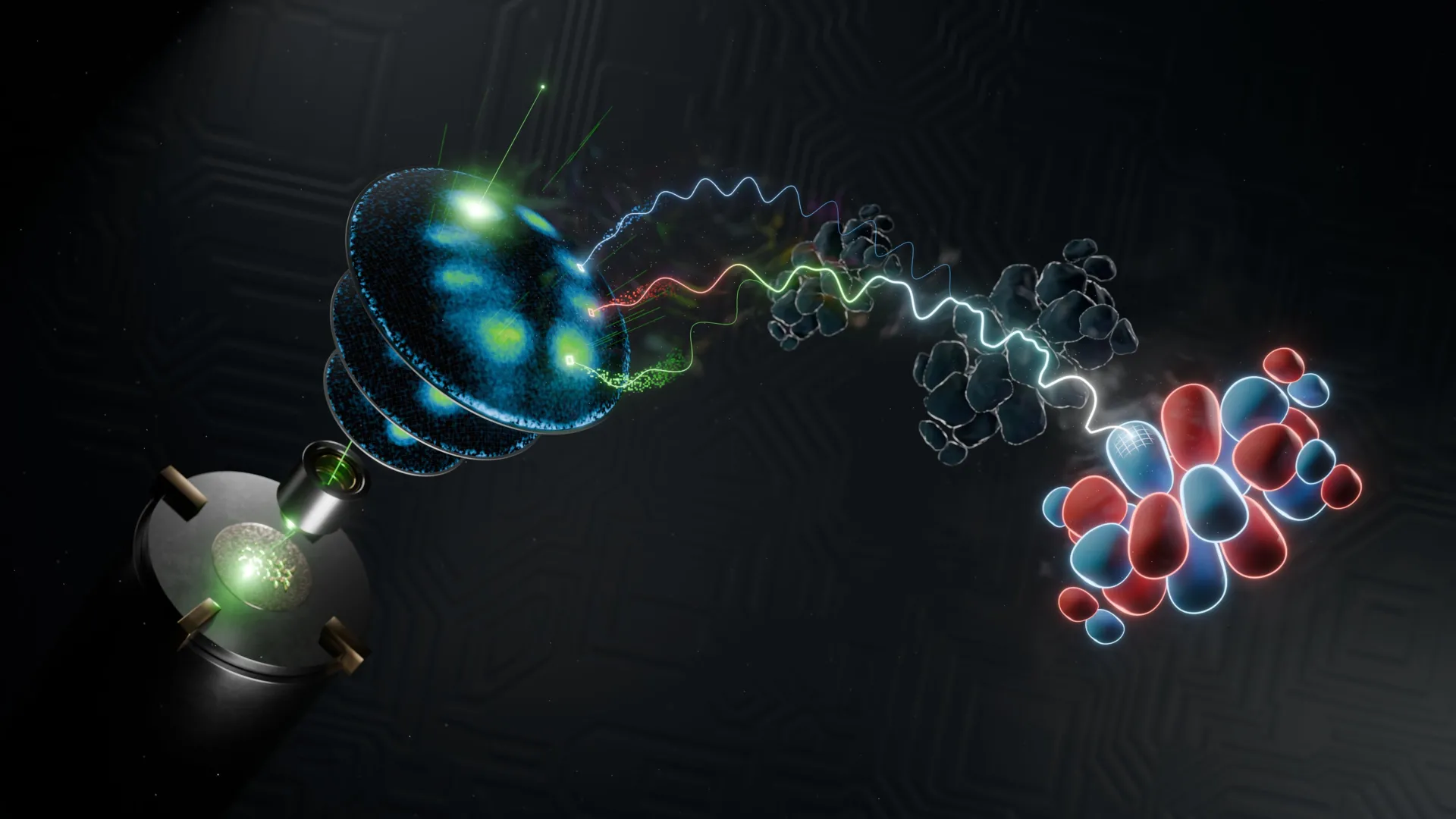
Quantum mechanics challenges the familiar idea that every object has one exact position at a given moment. Instead, fundamental particles such as electrons are described by a wave function—a mathematical expression that predicts the probability of finding properties such as position and momentum.
When an electron exists inside a molecule, its wave function is called a molecular orbital. Molecular orbitals reveal how molecules behave and interact with their surroundings, including how they absorb light and participate in chemical reactions. A complete three-dimensional image of an electron wave function could therefore provide valuable insight into molecular behavior. However, producing such images has remained a major scientific challenge.
Now, an interdisciplinary research team from the University of Göttingen has successfully reconstructed the three-dimensional wave functions of organic molecules measuring only a few nanometers across. The scientists combined advanced photoelectron spectroscopy with sophisticated mathematical algorithms to achieve the breakthrough, which is reported in Nature Communications.
Reconstructing the elusive quantum wave function
“The wave function is a fundamental quantity in quantum mechanics, but it cannot be directly observed or measured,” explains Professor Stefan Mathias of the University of Göttingen.
To overcome this challenge, the researchers used an indirect method known as photoelectron spectroscopy. The technique measures the momentum of electrons released from a molecule, providing access to important information about its wave function without directly disturbing its quantum state.
The team reconstructed the remaining information with advanced computer algorithms. This process enabled the researchers to create complete three-dimensional images of molecular orbitals and identify details smaller than the distance between carbon atoms within a molecule.
Previously, applying this approach in three dimensions required lengthy measurements at large synchrotron radiation facilities. That limitation made the method difficult to use on a broader scale and created a significant obstacle to imaging dynamic wave functions as three-dimensional videos at the atomic level.
Working toward ultrafast 3D molecular videos
Study co-leader Dr Matthijs Janssen of the University of Göttingen highlights the distinctive nature of the new method: “We are introducing two powerful new concepts. First, by fundamentally redesigning the computer algorithms, we can now obtain reliable 3D images from less experimental data. Second, the experiment uses a powerful laboratory-based soft X-ray source that generates ultrashort light pulses. It is the combination of these two technologies that makes this achievement possible.”
The improved algorithm substantially reduces the amount of experimental data needed to produce accurate three-dimensional wave-function images. At the same time, the laboratory-based soft X-ray source allows researchers to conduct experiments with extremely short light pulses. Together, these advances could make 3D molecular wave-function imaging faster, more accessible, and more practical.
Dr Wibke Bennecke, lead author of the study, said: “This technology could make stroboscopic videography a reality. It would allow us not only to observe the shape of a wave function, but also to track how it changes over femtoseconds. We could watch molecules respond to optical, electronic, or chemical changes with ultrafast temporal resolution and discover new ways to control these interactions at the scale of a few atoms.”
Further development of the technique could enable scientists to move beyond static molecular images and observe wave functions as they evolve in real time. Tracking these changes over femtosecond time scales may reveal how molecules respond to light, electronic forces, and chemical reactions, opening new possibilities for understanding and controlling interactions at the atomic scale.
Source: www.sciencedaily.com