


RSV F Protein Deep Mutational Scanning: Biosafety, Library Design and Antibody-Resistance Analysis
Biosafety and biosecurity
All experiments using pseudotyped lentiviral particles were conducted at biosafety level 2 (ref. 59). The pseudoviruses encoded only the RSV F protein and could therefore complete a single round of cell entry. They were not fully replicative infectious agents capable of causing disease.
The additional viral proteins required to produce the pseudotyped lentiviral particles—RSV G and the lentiviral Gag/Pol, Tat and Rev proteins—were supplied during pseudovirus production by transfection of four separate helper plasmids. These proteins were not encoded in the viral genome. Consequently, this study did not generate mutants of fully replicative biological agents capable of causing disease.
This study quantified how every single mutation in RSV F affects neutralization by antibodies, including antibodies used clinically. RSV is already evolving under widespread antibody-mediated selection in humans, and resistant strains are regularly identified in breakthrough infections3,4,6. Previously, escape mutations have been identified through breakthrough infections or by passaging authentic RSV in the presence of antibodies3,4,5,6,14,22,26.
Here, mutation effects were measured systematically outside the context of pathogenic virus. The resulting data support surveillance of ongoing RSV evolution affecting clinical antibodies and can inform the design of new antibodies that are more resilient to viral resistance.
RSV monoclonal antibodies
The RSV monoclonal antibodies nirsevimab13,19, clesrovimab14,40, palivizumab47, suptavimab2, RSM01 (ref. 11), 1A2 (ref. 52) and 1B6 (ref. 52) were produced by GenScript as human IgG1 kappa isotypes and Fabs.
Antibody sequences were obtained from the referenced publications, Protein Data Bank structures or original patents. The palivizumab sequence originated from patent US6955717B2. Antibody amino acid sequences are available in the RSV Long F deep mutational scanning repository. The Gates Medical Research Institute provided the RSM01 sequence.
Plasmids and primers
All plasmid and primer sequences used in this study are available through the study GitHub repository. Primers were obtained from Integrated DNA Technologies.
Cell lines and culture conditions
The cell lines used were 293T, sourced from the American Type Culture Collection, 293T-TIM1 (ref. 31) and Takara Lenti-X 293T. All cell lines were cultured in D10 medium consisting of Dulbecco’s Modified Eagle Medium supplemented with 10% heat-inactivated fetal bovine serum, 2 mM l-glutamine, 100 U ml−1 penicillin and 100 mg ml−1 streptomycin. Cells were maintained at 37 °C with 5% CO2.
Design of RSV F deep mutational scanning libraries
We created pseudovirus libraries containing nearly every possible single amino acid mutation in the ectodomain of the RSV F protein. The unmutated parental F sequence came from the Long strain of RSV, a subtype A, laboratory-adapted strain isolated in the 1950s (ref. 32).
The RSV F sequence was codon optimized using GenScript’s GenSmart codon optimization tool. Four amino acids were removed from the cytoplasmic tail to increase pseudovirus titres31. The lentiviral backbone contained an extended Gag sequence to improve lentiviral genome packaging, as previously described in refs. 60,61. The plasmid map for the lentiviral backbone containing the codon-optimized RSV F sequence is available on GitHub.
The library was designed to include all single amino acid substitutions in the RSV F ectodomain, spanning residues 26–529. This represented 504 × 19 = 9,576 possible mutations. Thirty stop codons, positioned at alternating sites from the beginning of the ectodomain, were also included as negative controls for cell-entry measurements.
A site-saturation variant library meeting these criteria was ordered from Twist Biosciences. The final Twist quality-control report is available as a CSV file on GitHub.
Cloning the RSV F deep mutational scanning plasmid libraries
Although the RSV F library was designed to contain all ectodomain mutations, 268 mutations were missing from the library produced by Twist Biosciences. We added these missing mutations and increased the representation of mutations in the nirsevimab-binding site, covering amino acid residues 62–69 and 196–212. This ensured that all possible mutations in the epitope were included and measured.
We generated a “spike-in” plasmid library using a mutagenesis PCR protocol62,63,64. NNS primers were designed for the nirsevimab-targeting sites using CodonTilingPrimers (https://github.com/jbloomlab/CodonTilingPrimers). Here, N represents any of the four nucleotide bases and S represents cytosine or guanine. NNS codons represent 32 codons encoding all 20 amino acids and a stop codon. Primers for the missing mutations were designed with TargetedTilingPrimers (https://github.com/jbloomlab/TargetedTilingPrimers).
Forward and reverse primer pools were prepared by combining the relevant NNS or targeted-mutation primers at an equal molar ratio per codon, producing a final concentration of 5 µM. Linear RSV F template was generated by digesting the lentiviral backbone containing codon-optimized RSV F with NotI-HF and NdeI. Mutagenesis PCR was then performed as described in ref. 30, except that seven PCR cycles were used to reduce the number of resulting multi-mutants. Remaining template was removed by digesting the PCR product with DpnI for 20 min at 37 °C. The spike-in mutagenesis PCR was performed in duplicate to generate libraries A and B.
The Twist Biosciences library pools were barcoded independently in two reactions to generate libraries A and B. These biological replicates were kept separate during all subsequent experimental steps. Barcoding followed ref. 30 and used primers containing a random 16-nucleotide sequence downstream of the RSV F stop codon. The only change from the published method was the use of 5 ng of template. The two spike-in mutagenized RSV F pools were also barcoded separately, resulting in four barcoding PCR reactions.
The lentiviral backbone (4016_V5LP_pHrU3_ForInd-Extgag_mcherry)61 was digested with MluI-HF and XbaI at 37 °C for 45 min, gel purified and further purified with AMPure XP beads (Beckman Coulter, A63881). Barcoded libraries were cloned into the lentiviral backbone at a 1:2 insert-to-vector ratio using a 1 h HiFi assembly at 50 °C. The assembly product was purified with AMPure XP beads and eluted in molecular-grade water.
Purified products were transformed into 10-beta electrocompetent cells (New England Biolabs, C3020K) with a Bio-Rad MicroPulser Electroporator using 2 kV for 5 ms. Ten electroporation reactions were performed for each barcoded Twist library and two reactions for each mutagenized spike-in library. After 1 h of recovery at 37 °C, transformed cells were pelleted from SOC medium, pooled and cultured overnight at 37 °C with shaking. Each Twist library was cultured in 150 ml of Luria–Bertani medium and each spike-in library in 100 ml, with ampicillin. Plasmids were extracted using the QIAGEN HiSpeed Plasmid Maxi Kit (QIAGEN, 12662).
Corresponding Twist and spike-in plasmid-library replicates were combined at a 1:1.25 Twist-to-spike-in molar ratio per codon. Long-read PacBio sequencing of the plasmid libraries showed that this ratio produced the most even distribution of mutants in the combined libraries.
Production of cell-stored RSV F deep mutational scanning libraries
Cells containing the deep mutational scanning libraries as single integrated RSV F-containing genomes were produced as described in ref. 30, with the changes summarized below and in Extended Data Fig. 1b.
VSV-G-pseudotyped lentiviruses were produced by transfecting four 10-cm dishes of 293T cells for each library. Each dish received 5 μg of the lentiviral backbone containing the barcoded RSV F library, 1.25 μg of each lentiviral Gag/Pol, Tat and Rev helper plasmid, and 1.25 μg of VSV-G expression plasmid. The helper plasmids were HDM-tat1b, pRC-CMV-Rev1b and HDM-Hgpm2 (Addgene product IDs 204154, 20413 and 204152)30; the VSV-G plasmid was pMD2.G (Addgene product ID 12259). BioT transfection reagent (Bioland Scientific, B01-02) was used.
Previous deep mutational scanning studies integrated libraries into a specific 293T-rtTA clone that produced high titres of pseudovirus for other viral entry proteins30. However, this clone did not produce high-titre RSV pseudovirus. After testing multiple 293T-rtTA clones, the highest titre from singly integrated cells was obtained with Takara’s Lenti-X 293T Cell Line (632180). This clonal 293T line does not overexpress rtTA.
The lentiviral backbone contained a doxycycline-inducible promoter. In other studies using 293T-rtTA cells and a similar backbone, doxycycline was added to induce expression from the integrated lentiviral backbone30. For the RSV F cell-stored libraries, titres were sufficiently high when expression was induced only by Tat during transfection. Transfecting rtTA and adding doxycycline provided no additional benefit, so the Takara Lenti-X 293T Cell Line was used.
To create the cell-stored library, VSV-G-pseudotyped viruses were used to infect the Takara Lenti-X 293T Cell Line at an infection rate below 1%. This rate was selected so that most transduced cells would receive only one integrated genome. Transduced cells were selected with puromycin, producing a final cell population in which each cell contained an integrated genome encoding one barcoded RSV F variant. Cells were expanded, frozen at a minimum of 2 × 10⁷ cells per aliquot and stored in liquid nitrogen.
Rescue of RSV F- and VSV-G-expressing pseudovirus libraries
Rescue of RSV F-expressing pseudoviruses
To rescue RSV F-expressing pseudoviruses from the integrated cells, 100 million cells were plated in five-layer flasks. The following day, each flask was transfected with 118.75 μg of each helper plasmid—HDM-tat1b, pRC-CMV-Rev1b and HDM-Hgpm2 (Addgene product IDs 204154, 20413 and 204152)—and 18.75 μg of the RSV G expression plasmid HDM_RSV_Long_G_31AACTdel (Addgene product ID 237350).
Xfect transfection reagent (631318) was used according to the manufacturer’s instructions, with 112.5 μl of reagent and 7.5 ml of buffer per five-layer flask. At 48 h post-transfection, supernatants were filtered through a 0.45-μm SFCA Nalgene 500-ml Rapid-Flow filter unit (09-740-44B).
Filtered supernatants were concentrated by ultracentrifugation through a 20% sucrose cushion in Hank’s buffered saline solution (Fisher, 14025092) at 100,000g for 1 h at above 20 °C. After the supernatant was discarded, pseudovirus pellets were resuspended in D10 medium. Concentrated F-expressing pseudoviruses were flash frozen as described in ref. 31 and stored at −80 °C for downstream selection experiments.
Rescue of VSV-G-expressing pseudoviruses
To rescue VSV-G-expressing pseudoviruses, 100 million integrated cells were plated in five-layer flasks. The following day, each flask was transfected with 37.5 μg of each helper plasmid—HDM-tat1b, pRC-CMV-Rev1b and HDM-Hgpm2—and 37.5 μg of the VSV-G expression plasmid pMD2.G (Addgene product ID 12259).
BioT transfection reagent was used according to the manufacturer’s instructions. At 48 h post-transfection, supernatants were filtered through a 0.45-μm SFCA Nalgene 500-ml Rapid-Flow filter unit and concentrated with Lenti-X concentrator (Takara, 631232) at a 1:3 virus-to-concentrator ratio. Samples were incubated at 4 °C for 3 h and centrifuged at 1,500g at 4 °C for 45 min. The supernatant was discarded, and viral pellets were resuspended in D10 medium and stored at −80 °C.
PacBio long-read sequencing for RSV F variant–barcode linkage
Long-read PacBio sequencing was used to link RSV F mutations to their barcode sequences in lentiviral genomes from pseudoviruses produced by the cell-stored libraries. Barcode–mutation linkage was measured after generating the singly integrated cell libraries because template switching between the pseudodiploid lentiviral genomes during reverse transcription can change barcode–variant pairings relative to the original plasmid pool. Sequencing the integrated library ensured that the linkage reflected the actual integrated provirus.
The overall process followed ref. 30, with the following modifications. A total of 1 × 10⁶ 293T-TIM1 cells were plated in each well of six-well plates coated with poly-l-lysine. The next day, cells were infected with 30 million transducing units (TU) of VSV-G-expressing library pseudoviruses, using six wells per library and 5 million TU per well.
At 12 h post-infection, non-integrated reverse-transcribed lentiviral genomes were recovered by miniprepping the 293T-TIM1 cells as described in ref. 30. Amplicons for long-read sequencing were prepared using the approach described in refs. 30,64. PCR products for each library were combined, and amplicon length was verified with a TapeStation before sequencing. Each library was sequenced on a single-molecule real-time sequencing cell with a 30 h video time on a PacBio Sequel IIe sequencer. Each library was sequenced a second time to maximize identification of variants.
Sequencing data were processed with the dms-vep-pipeline-3 package. PacBio circular consensus sequences were aligned to the unmutated RSV F reference sequence using the alignparse package65. Reads were excluded if they aligned poorly, contained more mutations than expected in unmutated regions, lacked a barcode or resulted from strand exchange.
Consensus sequences for each barcode and/or variant sequence were generated with alignparse. The analysis required at least three circular consensus sequence (CCS) reads and used a maximum cut-off of 0.2 for minor variants within a consensus. Final barcode–variant lookup tables were used as references for all subsequent Illumina short-read sequencing of barcodes.
The final barcode–variant tables are available in the study GitHub repository.
The final libraries contained 57,888 unique barcoded variants for LibA and 64,078 for LibB. These libraries covered 99% and 99.4% of all amino acid mutations, respectively (Extended Data Fig. 1c). More than half of the variants contained a single amino acid mutation, while the remaining variants contained zero or multiple mutations (Extended Data Fig. 1d).
Measuring the effects of RSV F mutations on cell entry
We measured the effects of RSV F mutations on cell entry using the approach described in ref. 30, with the modifications below. 293T-TIM1 cells were infected with pseudovirus libraries displaying RSV F mutants. In parallel, cells were infected with control VSV-G pseudovirus, whose entry is independent of RSV F function (Extended Data Fig. 2a).
A total of 2 × 10⁶ 293T-TIM1 cells were plated in each well of six-well plates coated with poly-l-lysine. The next day, cells were infected with approximately 12 × 106 TU in total of the F-pseudovirus library, using 3 × 10⁶ TU per well, or approximately 3 × 10⁷ TU in total of the VSV-G pseudovirus library, using 5 × 10⁶ TU per well.
After F-pseudovirus addition, cells were spun at 900g for 3 h at 30 °C. In some selections, 20 μg ml−1 diethylaminoethyl (DEAE)-dextran was added during infection because it increased pseudovirus titres. However, poor cell health reduced recovery of infecting barcodes, so DEAE-dextran was not used in follow-up selections.
At 12 h post-infection, non-integrated reverse-transcribed lentiviral genomes were recovered by miniprepping the cells. Amplicons for dual-indexed Illumina sequencing were prepared as described in ref. 64. Samples were pooled in equal DNA amounts, separated on a 1% agarose gel, and the correct-size band was excised and purified with AMPure XP beads. Samples were diluted to 5 nM and sequenced on an Illumina NextSeq 2000 with a P3 reagent kit or on a NovaSeq X Plus system.
Mutation effects were quantified as the log2 cell-entry effect relative to unmutated RSV F. Single- and multi-mutant data were jointly analysed with global-epistasis models33,34. Illumina reads were aligned to the barcode–variant table generated from the PacBio CCS data. Barcode frequencies were then compared between the VSV-G and F-mutant selections using the dms_variants package, as previously described in ref. 30.
Cell-entry scores were calculated using the log-enrichment ratio:
log2 [(nvpost/nwtpost)/(nvpre/nwtpre)]
Here, nvpost is the count of variant v in the F-pseudotyped infection, or post-selection condition; nvpre is the count of variant v in the VSV-G-pseudotyped infection, or pre-selection condition; and nwtpost and nwtpre are the corresponding counts for wild-type variants.
Positive cell-entry scores indicate that a variant entered cells more efficiently than the unmutated parental F, whereas negative scores indicate poorer entry. As expected, variants with only synonymous mutations had wild-type-like scores of zero. Stop-codon variants had strongly negative scores, while amino acid variants ranged from wild-type-like to highly impaired (Extended Data Fig. 2b).
Mutation-level cell-entry effects were calculated by fitting a sigmoid global-epistasis function to variant entry scores after truncating values at a lower bound corresponding to the median functional score of all stop-codon variants. The multi-dms software package was used for this analysis. A mutation effect of zero indicates no effect on entry; negative values indicate impaired entry; and positive values indicate improved entry.
Final mutation-level entry effects were calculated from three technical replicates for each library, producing six functional selections in total—three from LibA and three from LibB. Mutations were required to occur with at least two unique barcodes. Mutations with high standard deviations between replicates were removed. Entry effects were highly correlated between replicates and libraries (Extended Data Fig. 2c,d). Values shown in the figures represent the average across libraries and replicates.
Measuring RSV F mutation effects on antibody neutralization
Antibody-neutralization effects were measured using the method described in ref. 30, with the modifications below. A total of 2 × 10⁶ 293T-TIM1 cells were plated in each well of six-well plates coated with poly-l-lysine. The following day, approximately 1.5–2 × 10⁶ TU of the F-pseudovirus library were incubated with D10 medium alone as a no-antibody control or with antibody for 1 h before being added to the cells.
For monoclonal antibodies, incubations were performed in a total volume of 2 ml to reduce the possibility of ligand depletion because these antibodies have very high potency. Antibody concentrations were selected to span a range in which approximately 50% to 99.5% of variants were neutralized.
During DNA template extraction, a plasmid containing eight known barcodes was added at a level expected to represent approximately 1% of reads in the no-antibody control. This DNA spike-in standard enabled estimation of neutralization in each antibody condition relative to the no-antibody control, as described in refs. 30,64 and shown in Extended Data Fig. 4a.
After Illumina barcode sequencing, the fraction of infectivity retained at each antibody concentration was calculated from the DNA-standard barcode counts. The polyclonal software66 was then used to fit neutralization curves and estimate the effects of mutations on antibody neutralization.
Mutation effects were retained only when mutations had at least two unique barcodes. Mutations with very low cell-entry scores or high standard deviations across neutralization replicates were excluded. Reported effects represent averages across all replicates, with at least two independent experimental selections from each of the two libraries, LibA and LibB.
Validation of RSV F cell-entry effects with individual pseudoviruses
To validate cell-entry effects, we generated plasmids expressing individual RSV F mutations in a lentiviral backbone. The parental RSV F amino acid sequence was identical to the unmutated sequence used in the deep mutational scanning lentiviral vector.
Selected mutations covered a range of measured entry effects: N67I, S215V, S215P, S398L, E87N and D486N. These constructs were transfected into 293T cells with the helper plasmids HDM-tat1b, pRC-CMV-Rev1b and HDM-Hgpm2 (Addgene product IDs 204154, 20413 and 204152)30, together with RSV G in the HDM_RSV_Long_G_31AACTdel expression plasmid (Addgene product ID 237350).
After 48 h, supernatants were filtered through 0.45-μm filters to remove cell debris. Pseudoviruses were titrated on 293T-TIM1 cells, and transducing units per millilitre were measured by flow cytometry as described in ref. 31. Average titres from two replicates for each pseudovirus were compared with the average titre of unmutated RSV F.
Cell-entry effects of amino acid differences in natural RSV sequences
An alignment of RSV F-protein sequences from subtypes A and B was generated on 27 October 2025 using the RSV Nextstrain workflow45. Amino acid differences were identified relative to the laboratory-adapted subtype A Long strain used for deep mutational scanning.
These differences were used to compare the distribution of cell-entry effects for mutations observed in natural sequences with the distribution for all mutations measured by deep mutational scanning. The percentage of natural sequences containing each mutation was also calculated, as shown in Extended Data Fig. 3. Additional details are available in the sequence-variation analysis notebooks.
Validation of RSV F mutations affecting antibody neutralization
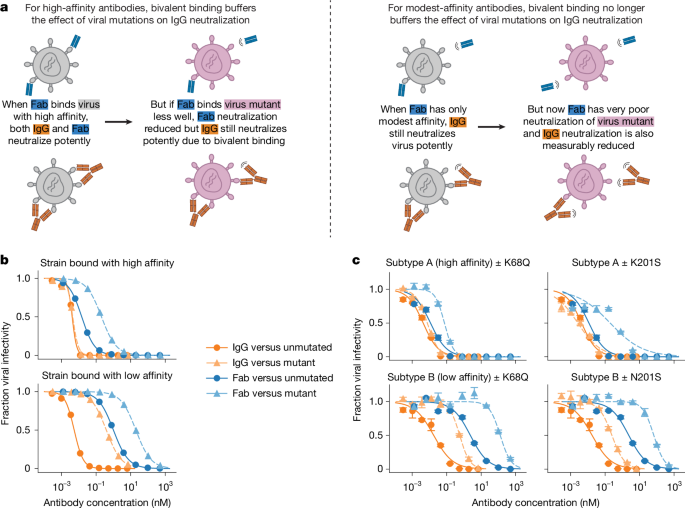
Previously known and newly identified resistance mutations were selected for nirsevimab and clesrovimab across a range of measured neutralization effects. The mutations were used to assess the effects of subtype A and B sequence backgrounds on neutralization by antibody IgG and Fab and to validate deep mutational scanning measurements.
Single amino acid mutations were introduced into subtype A and subtype B expression vectors. Subtype A used the Long strain, which was also used for deep mutational scanning, and subtype B used the B1 strain. Plasmids were generated by Twist Biosciences. Pseudoviruses were produced and neutralization was measured as described in ref. 31.
All F constructs included the complete cytoplasmic tail and were paired with RSV G in the HDM_RSV_Long_G_31AACTdel expression plasmid (Addgene product ID 237350). Plasmid maps are available in the study GitHub repository. In all plotted neutralization curves, points represent the mean and standard error of at least two replicate measurements.
Nirsevimab resistance mutations
Subtype A mutations affecting nirsevimab neutralization were N67T, K68N, K68Q, D73N, K201S, K201T, P205S, V207E, K209D, K209Q, Q210T, S211R and S215K. Subtype B mutations were K68N, K68Q, D73N, N201S, N201T, P205S, Q209D, Q210T, S211R and S215K. V207E did not produce usable pseudovirus titres in the B1 background. These results are shown in Fig. 3 and Extended Data Fig. 4.
T67N and Q209K were also generated in subtype B. These mutations restored the amino acids found at the corresponding positions in the subtype A Long strain. Results for these B1 mutants, together with the corresponding subtype A mutations N67T and K209Q, are shown in Extended Data Fig. 4. Subtype A K68Q and K201S and subtype B K68Q and N201S are shown in Figs. 1 and 3 and Extended Data Fig. 4.
The Long and B1 wild-type neutralization curves shown in Fig. 3d,f are representative. All replicates are available in the validation analyses repository. Correlation plots in Figs. 3 and 4 show fold-change IC50 values relative to wild type. Each mutation was compared with a matched wild-type measurement from the same experimental date, while the wild-type point in the correlation plots represents the geometric mean IC50 of all wild-type replicates.
Clesrovimab resistance mutations
Mutations affecting clesrovimab neutralization in subtypes A and B were R429M, R429S, S443P and G446D. These results are shown in Fig. 4 and Extended Data Fig. 6.
The F sequences called “A2020” and “B2024” in Extended Data Fig. 6a were taken from natural strains broadly representative of recent subtype A and B strains31. Their GenBank accession numbers are PP495954.1 and PP660445.1, respectively.
Interactive Nextstrain trees with RSV antibody-escape scores
Computer code was integrated into the Nextstrain RSV view to score natural RSV sequences for antibody resistance and display the results on phylogenetic trees. The implementation is available in the Nextstrain RSV GitHub repository.
The existing Nextstrain view downloaded more than 60,000 RSV sequences from Pathoplexus46 and constructed separate subtype A and B phylogenetic trees. Each tree was subsampled to approximately 3,000 sequences selected to represent different time periods and countries.
We added code that assigns each sequence an escape score derived from the deep mutational scanning data. Scores were calculated either as the sum of the effects of all constituent mutations or as the maximum effect of any mutation on neutralization by the Fab or IgG form of nirsevimab or clesrovimab.
Interactive trees can be coloured by escape score using the “Color By” option in the left toolbar. An example is available at https://nextstrain.org/rsv/b/F-antibody-escape/6y?c=Nirsevimab-Fab_total_escape. The interface also provides an option to label sequences by their top escape mutation.
Escape-score calculations were integrated into Nextstrain RSV builds for both F sequences and full genomes. Builds are available for all time periods, the past six years and the past three years. Because the builds are updated with the latest available sequences, the linked views display the current sequence data.
We also created “F-antibody-escape” builds in the Nextstrain RSV views. These trees are subsampled to include all sequences with high predicted nirsevimab or clesrovimab escape scores and therefore over-represent resistant strains. They do not provide an accurate estimate of the prevalence of resistance mutations. However, they are preferred when the goal is to identify top resistant strains that might otherwise be removed during subsampling. The build can be selected using the “change dataset” option in the left toolbar.
Validation of RSV strains with predicted neutralization resistance
Natural RSV F sequences with predicted resistance to nirsevimab or clesrovimab were identified using the interactive Nextstrain phylogenetic trees with antibody-escape scores. Selected sequences were codon optimized and cloned into expression vectors by Twist Biosciences.
Sequences with predicted resistance to nirsevimab Fab or IgG were PP_002XVQT, PP_002KSRJ, PP_002QMLP, PP_002WHEU, PP_002WWH8, PP_002SUFP, PP_001QYN9, PP_001Y2UB and PP_003W55P. Sequences with predicted resistance to clesrovimab Fab or IgG were PP_002W1BG, PP_001WGC0, PP_001Y62S and PP_001ZQ7W.
PP_001W26S and PP_002UDSB were subtype A and B controls, respectively, also referred to as A2020 and B2024 in our previous study31. A complete list of key sequences is available in Pathoplexus under SeqSet PP_SS_628.1 (ref. 67). All sequences shown in Fig. 5 and Extended Data Fig. 8 are available under SeqSet PP_SS_661.1 (ref. 68) for subtype A and SeqSet PP_SS_662.1 (ref. 69) for subtype B.
All plasmid maps are available through the strain-validation directory on GitHub. The constructs were used to generate RSV pseudoviruses expressing F from the natural sequences, paired with the RSV G expression plasmid HDM_RSV_Long_G_31AACTdel (Addgene product ID 237350) as previously described in ref. 31.
Pairing all natural F sequences with the same RSV G construct prevented G-related variability from being introduced into the comparison. The resulting pseudoviruses were tested in neutralization assays with nirsevimab or clesrovimab IgG and Fab31.
To ensure reliable neutralization curves, the no-antibody virus-only control was required to produce at least 400,000 relative light units per well. For strains with titres near this threshold, additional controls were included to determine whether apparent escape was specific to the monoclonal antibody rather than an experimental artefact.
Structural analysis of RSV F antibody-binding sites
UCSF ChimeraX70 was used for structural visualization. All Protein Data Bank accession identifiers used in the analysis are provided in the figure legends.
Reporting summary
Additional information on the research design is available in the Nature Portfolio Reporting Summary linked to this article.
Source: www.nature.com