


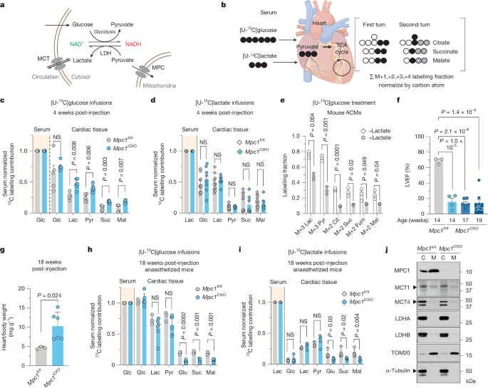
Researchers at the University of Utah studied how cardiac metabolism supports heart function using human myocardial samples, genetically modified mice, cardiac injury models, isotope tracing, mitochondrial assays and imaging. The work included samples from patients with advanced heart failure and non-failing organ donors, alongside mouse models lacking the monocarboxylate transporter MCT1, MCT4 or mitochondrial pyruvate carrier 1 in cardiac tissue. The investigators assessed cardiac performance, exercise capacity, ischemia–reperfusion injury, metabolism, mitochondrial respiration, protein localization and structural changes using biochemical, imaging and statistical methods approved by the relevant ethics committees.
Human heart samples
Human myocardial tissue was obtained through the Utah Cardiac Recovery Program at the University of Utah under an approved Institutional Review Board protocol (IRB-00030622). All samples were collected with informed consent from patients, organ donors or their guardians.
Samples from consented patients were collected at the time of heart transplantation after chronic, advanced heart failure with reduced ejection fraction. Non-failing donor hearts that could not be used for transplantation for non-cardiac reasons, such as donor–recipient size mismatch or infection, served as controls. Myocardial tissue was collected in the operating room, immediately frozen in liquid nitrogen and stored at −80 °C until use.
Animal care and mouse models
All animal procedures were approved by the University of Utah Institutional Animal Care and Use Committee (IACUC). Mice were housed at 22–23 °C on a 12 h light:12 h dark cycle. They received irradiated chow (Teklad Diet 2920x) and unlimited access to water. Food was withdrawn only during experiments, beginning at the start of the light cycle.
Adult mice with cardiac-specific deletion of MPC1 (Mpc1fl/fl; αMHCMerCreMer) were generated as previously described51. Mct1fl/fl mice were generated as previously described52. Cardiac-specific Cre recombinase was introduced by crossing the mice with αMHCMerCreMer mice from Jackson Laboratories strain 005657.
Eight-week-old Mct1fl/fl:αMHCMerCreMer+/− mice and Mct1fl/fl;αMHCMerCreMer−/− littermates received intraperitoneal tamoxifen injections at 40 mg kg−1 for three consecutive days. This produced adult inducible cardiac-specific Mct1 knockout mice (Mct1iCKO) and Mct1fl/fl control littermates.
Adult cardiac-specific MCT4-knockout mice (Mct4fl/fl;αMHC-MerCreMer) were generated from three embryonic stem-cell clones carrying Slc16a3 tm1a, which encodes MCT4. The clones were obtained from the European Conditional Mouse Mutagenesis program/European Mouse Mutant Archive (EUCOMM/EMMA; http://www.infrafrontier.eu/). All clones came from the JM8A3.N1 cell line, which has a C57Bl/6NTac background and agouti coat-colour distribution.
The embryonic stem-cell clones were injected into the blastocoel of 3.5-day-old mouse blastocysts from the C57Bl/6 Tyrc-Brd host strain. The injected embryos were transferred to the uterine horns of appropriately timed pseudopregnant C57BL/6-Tyrc-Brd females. Chimeras with the greatest embryonic stem-cell contribution were bred with C57Bl/6J mice to test for germline transmission of the targeted allele. Pups carrying germline-transmitted tm1a were identified and propagated. This first allele was a reporter-tagged knockout insertion with conditional potential. Mice carrying the appropriate floxed alleles were crossed with αMHCMerCreMer mice as described above. Experiments began four weeks after injection, when the mice were 12 weeks old.
Cardiac function and disease models
Echocardiography
Mice were anaesthetized with 1.5% isoflurane (Vet One, NDC 13985-046-60). Two-dimensional long-axis and short-axis echocardiographic images were captured sequentially using a Vevo imaging system (VisualSonics) and analysed with Vevo Strain software version 3.1.1. Measurements were based on three consecutive cardiac cycles. Limb leads were attached with conductive cream to record an electrocardiogram.
Angiotensin II and phenylephrine infusion
Twelve-week-old C57BL/6 mice were divided into four groups, weighed and screened by echocardiography before surgery to establish baseline cardiac parameters. Surgical instruments were sterilized with a glass-bead sterilizer, and aseptic technique was used.
Alzet osmotic minipumps (model 2006) were filled with angiotensin II at 1.5 μg g−1 day−1 and phenylephrine at 50 μg g−1 day−1 in 0.9% NaCl. The pumps delivered the solution at 0.15 μl h−1 for 42 days, according to the manufacturer’s instructions. They were implanted subcutaneously on the back through a small incision while the mice were under 1.5% isoflurane in oxygen.
After implantation, the incision was closed and the mice were placed in a clean, warm cage and monitored for distress. Mice were weighed and assessed weekly by echocardiography. After 42 days of angiotensin II/phenylephrine treatment, they were euthanized by cervical dislocation, and blood and heart tissue were collected.
Transverse aortic constriction
Baseline echocardiograms were obtained before transverse aortic constriction (TAC) surgery in male and female Mct1fl/fl and Mct1iCKO mice aged 12–14 weeks, using previously described methods53,54. Mice received sustained-release buprenorphine SR subcutaneously at 0.15 mg kg−1 before surgery. Anaesthesia was maintained with 2–3% isoflurane in oxygen delivered through a nose cone connected to a VetFlo vaporizer.
The aortic arch was exposed and constricted with a titanium clip. The incision was closed in layers with 6-0 sutures. During recovery, regular-release buprenorphine was administered as needed for up to 48 h. Unclipped Mct1fl/fl and Mct1iCKO mice served as sham controls; they underwent anaesthesia and a small incision but no aortic constriction or cardiac injury. Echocardiography was performed weekly for six weeks after surgery to assess cardiac structure and function.
Ischemia–reperfusion
Ischemia–reperfusion experiments used 20–30 g, 12–14-week-old Mct1iCKO and Mct1fl/fl mice four weeks after tamoxifen administration and before the onset of heart-failure symptoms. Mice were anaesthetized with 2–3% isoflurane, intubated with a 22 G intravenous catheter and ventilated at 100 breaths per minute with a small-animal respirator. They were placed supine on a warming pad at 38 °C. Chest hair was removed with depilatory cream, which was rinsed off with water or saline to prevent skin irritation. The skin was prepared with betadine and alcohol.
A thoracotomy was performed and a retractor was used to improve visibility. The pericardium was carefully removed without damaging the myocardium. Under a dissecting microscope, the left anterior descending coronary artery (LAD) was identified and a 6-0 silk suture was placed beneath it. A loose double knot with a 2–3 mm loop was formed, and a 2–3 mm piece of PE-10 tubing was placed through the loop. The tubing had been soaked in 100% ethanol and rinsed with sterile water. Tightening the loop around the artery produced 30 min of coronary occlusion.
Successful LAD ligation was confirmed by a paler colour in the anterior wall of the left ventricle. After 30 min, the knot was released and the tubing removed. Reperfusion was confirmed by the return of a red colour to the ventricular wall and continued for 2 h.
For acute experiments, the artery was briefly re-occluded after injury and Evans blue dye was injected into the left atrium to identify the area at risk. Hearts were removed, perfused with saline, weighed and cut into 1 mm slices with a vibratome. Alternatively, ischemic and non-ischemic regions were separated and flash-frozen in liquid nitrogen. Slices were incubated in 1% triphenyltetrazolium chloride at 38 °C for 20 min and then fixed in 10% formalin to distinguish viable from necrotic tissue. Sections were imaged the following day, and myocardial salvage was quantified in ImageJ by researchers blinded to the experimental groups.
For chronic experiments, mice underwent 30 min of ischemia, after which the ligature was removed and the incisions were sutured closed. Mice were placed in a warm cage, monitored for distress, weighed and assessed weekly by echocardiography.
Exercise capacity
Twelve- to 14-week-old Mct1fl/fl and Mct1iCKO mice completed a five-day treadmill-acclimation protocol before maximal exercise testing. The protocol familiarized the mice with the procedure without producing a training effect55.
After a day of rest, mice were weighed and tested on a treadmill. The test began with a 10 min warm-up at 10 m min−1 and a 10% incline. Speed then increased by 5 m min−1 every 2 min. The test ended when a mouse either resisted a physical stimulus, such as touching the tail or hindquarters with a bristle brush, for more than 20 s or remained on the stationary rear portion of the belt five times within 30 s. The stopping time was recorded, and the mouse was returned to its cage to recover. Maximum speed did not exceed 30 m min−1. Total work, measured in kg m−1, and time to exhaustion, measured in minutes, were calculated. Blood lactate was measured at 10, 20 and 30 m min−1 at a 10% incline to assess systemic effects of lactate production in Mct1iCKO mice relative to controls.
Catheterization and isotope tracing
Jugular-vein catheterization
Sterile mouse jugular-vein catheters were obtained from SAI Infusion Technologies, and a single-channel vascular-access button was obtained from Instech Laboratories. Mice were fasted before surgery, shaved and given bupivacaine before anaesthesia. A Leica S9i stereo dissection microscope was used during catheter placement. Mice received standard postoperative support and were allowed to recover for at least five days before further experiments. Animals with impaired vital signs, activity or reflexes were euthanized and excluded from experimental studies.
Stable-isotope infusions
Twelve-week-old mice with surgical catheters were maintained on a normal light cycle from 06:00 to 18:00. On the infusion day, they were moved to new cages without food at 06:00, the beginning of their sleep cycle. Metabolite-specific infusions lasted 2–3 h and began at approximately 12:00 to reach serum steady state, based on previously published studies.
The infusion system from Instech Laboratories used a swivel and tether that allowed the mice to move freely. Water-soluble isotope-labelled metabolites from Cambridge Isotope Laboratories were prepared in sterile normal saline. For 13C-labelled fatty acids, the fatty acids were complexed with bovine serum albumin at a 4:1 molar ratio.
Infusion rates were 0.1 μl min−1 g−1 for water-soluble metabolites and 0.4 μl min−1 g−1 for fatty acids. The solutions and infusion times were:
- 200 mM [U-13C]glucose for 3 h;
- 490 mM [U-13C]sodium lactate for 2 h;
- 50 mM [U-13C]3-hydroxybutyrate for 3 h;
- 8 mM [U-13C]oleic acid for 3 h; and
- 4 mM [U-13C]sodium palmitate for 3 h.
For [U-13C]glucose infusions with AZD treatment, mice received 100 mg kg−1 AZD or vehicle by oral gavage 6 h before the infusion and again when the infusion began. The vehicle contained 0.5% hydroxypropyl methylcellulose and 0.1% Polysorbate 80.
Blood was collected by tail snip, approximately 10 μl at a time, into Microvette CB 300 Z tubes. Samples were kept on ice and centrifuged at 5,000g for 5 min at 4 °C to obtain serum. At the end of the infusion, mice were euthanized by cervical dislocation. Tissues were rapidly dissected, rinsed in cold PBS, clamped with a pre-chilled Wollenberger clamp and dropped into liquid nitrogen.
Tail-vein infusion
Mice were anaesthetized with 2% inhaled isoflurane for the entire infusion. A 30 G catheter was inserted into the tail vein. For glucose infusion, 0.2 M [U-13C]glucose was administered as a bolus at 1.6 μl g−1 min−1 for 2 min, followed by a constant infusion of 0.1 μl g−1 min−1 for 30 min. For lactate infusion, 1.67% [U-13C]lactate was given as a bolus at 0.96 μl g−1 min−1 for 2 min, followed by a constant infusion of 0.3 μl g−1 min−1 for 30 min. Blood was collected retro-orbitally during the procedure, and tissues were harvested immediately after infusion as described for jugular-catheter experiments.
Metabolite extraction and mass spectrometry
Tissue and serum extraction
For serum extraction, 4 μl of serum was diluted with 140 μl of ice-cold 80% methanol/20% water and vortexed. Chloroform, 72 μl, was added, and the sample was vortexed and centrifuged at 15,060g for 10 min at 4 °C to separate the phases. The aqueous supernatant was used for liquid chromatography–mass spectrometry (LC–MS).
For tissue extraction, 30–40 mg of snap-frozen mouse tissue was placed in a pre-chilled Safe-Lock tube (Eppendorf, 022363352) containing a cold 5/16-inch stainless-steel ball (Grainger, 4RJL8). Tissue was disrupted at 25 Hz for 30 s under liquid nitrogen using a Retsch CryoMill (20.749.0001).
Frozen polar-metabolite extraction solution containing 40:40:20 acetonitrile:methanol:water and 0.1% formic acid was added at 15 μl per milligram of tissue. Samples were vortexed and neutralized with 8 μl of 15% ammonium bicarbonate per 100 ml of extraction solvent. They were centrifuged at 16,000g for 10 min at 4 °C. The pellet was extracted a second time with 525 μl of chilled 40:40:20 methanol:acetonitrile:water. The supernatants were combined, mixed with 525 μl of chloroform and centrifuged again at 16,000g for 10 min at 4 °C. The final aqueous layer was transferred to LC–MS tubes.
LC–MS analysis of polar metabolites
Polar metabolites were separated by hydrophilic interaction liquid chromatography using a Vanquish HPLC system (ThermoFisher Scientific) and an Xbridge BEH amide column (2.1 mm × 150 mm, 2.5 µm particles, 130 Å pores; Waters). The mobile-phase gradient used solvent A, consisting of 20 mM ammonium hydroxide and 20 mM ammonium acetate in 95:5 acetonitrile:water at pH 9.5, and solvent B, 100% acetonitrile. The flow rate was 150 μl min−1.
The gradient was: 0 min, 90% B; 2 min, 90% B; 3 min, 75% B; 7 min, 75% B; 8 min, 70% B; 9 min, 70% B; 10 min, 50% B; 12 min, 50% B; 13 min, 25% B; 14 min, 25% B; 16 min, 0% B; 20.5 min, 0% B; 21 min, 90% B; and 25 min, 90% B. The autosampler was maintained at 4 °C, the column at 30 °C and the injection volume was 3 μl.
Samples were introduced by electrospray ionization into a Q Exactive HF Orbitrap mass spectrometer (ThermoFisher Scientific) operated in negative-ion mode. The instrument had a resolving power of 75,000 at m/z 200 and scanned from 75 to 1,000. Data were collected with Xcalibur software and analysed with EL-MAVEN. Peaks were assigned using exact mass and comparisons with known standards56. Intensities were corrected for natural isotope abundance with the AccuCor R package57. Untargeted LC–MS data were analysed with MetaboAnalyst.
Tricarboxylic-acid-cycle labelling
Total carbon fractional contributions were calculated by dividing the natural-isotope-corrected experimental labelling fraction by the number of carbon atoms in each metabolite. This produced a single carbon-weighted labelling fraction for each metabolite. Values were normalized to the serum tracer-weighted labelling fraction, which was represented as 1 in all graphs38,58.
Heart perfusion and cardiomyocyte studies
Langendorff perfusion
Mice received 100 IU heparin intraperitoneally 20 min before anaesthesia with 3% isoflurane and cervical dislocation. Hearts were rapidly removed and cannulated on a Langendorff apparatus operating at 2.6 ml min−1.
The modified Krebs–Henseleit buffer contained 118 mM NaCl, 4.7 mM KCl, 2.5 mM CaCl2, 1.2 mM KH2PO4, 1.2 mM MgSO4, 24 mM NaHCO3, 0.5 mM EDTA, 0.7 mM oleic acid complexed to 2% albumin, 11 mM glucose, 50 μM L-carnitine, 1.5 mM lactate, 0.2 mM pyruvate, 0.8 nM insulin and 5 nM adrenaline. These conditions were intended to reproduce the non-fasted substrate and hormone state of mice as previously described1. The buffer was equilibrated with 95% O2/5% CO2 and maintained at 37 °C.
Hearts were retrogradely perfused in non-recirculating mode for 30 min while beating spontaneously. For isotope tracing, unlabelled substrates were replaced with [U-13C]glucose, [U-13C]lactate or [2-2H]lactate. Electrocardiogram leads connected to an AD Instruments PowerLab 8/35 were positioned on the cannula just above the aorta and on the apex. Heart rate was averaged over 30 s during the final minute of perfusion. Hearts were then clamped with a pre-chilled Wollenberger clamp and dropped into liquid nitrogen, or mitochondria were isolated as described below.
Adult cardiomyocyte isolation
Adult cardiomyocytes were isolated using established protocols16,38. Twelve-week-old mice were anaesthetized with isoflurane, after which the heart was removed and attached to an aortic cannula. The heart was perfused with oxygenated solutions at 37 °C and pH 7.3.
A calcium-free solution was perfused for 5 min, followed by 15 min with the same solution containing 1 mg ml−1 collagenase and 0.1 mg ml−1 protease. The heart was then perfused for 1 min with stopping solution containing 20% serum and 0.2 mM CaCl2. All perfusions used a flow rate of 2 ml min−1.
After the atria were removed, the ventricles were teased apart with forceps, gently rocked for 10 min and passed through a nylon mesh. After gravity sedimentation, cells were stored at 37 °C in normal HEPES-buffered solution before plating. The isolated myocytes had rod-shaped morphology with distinct striations and no spontaneous contractions.
Cardiomyocyte culture and growth assays
Adult cardiomyocytes were incubated with tracers in DMEM or human plasma-like medium (HPLM), as specified. HPLM was prepared in-house as previously described59. Glucose or lactate was selectively omitted and replaced with heavy-isotope tracers when required.
Plating media contained 100 μg ml−1 Primocin, 100 U ml−1 penicillin-streptomycin, 10 mM HEPES, 5% dialysed fetal bovine serum and 10 mM 2,3-butanedione monoxime. Isolated cardiomyocytes were enriched by gravity sedimentation in increasing calcium concentrations, plated on laminin-coated Petri dishes or coverslips and allowed to adhere for at least 1 h in a 5% CO2 incubator.
Cells were then transferred to HPLM- or DMEM-based medium containing Primocin, penicillin-streptomycin, HEPES, insulin-transferrin-selenium, 0.1 mg ml−1 BSA and a physiological BSA-conjugated fatty-acid mixture, as previously described60.
Permeabilized cardiomyocyte assays and imaging
Cardiomyocytes were isolated and cultured in HPLM without 2,3-butanedione monoxime. After adhering for at least 1 h to a laminin-coated 24-well plate, cells were washed with warm PBS and permeabilized for 5 min with 0, 25 or 100 μg ml−1 saponin in mitochondrial assay buffer (MAB). Cells were washed once with MAB and incubated at 37 °C.
MAB contained 2 mM KH2PO4, 120 mM KCl, 3 mM HEPES, 1 mM EGTA, 3 g l−1 essentially fatty-acid-free BSA, 1 mM lactate, 50 μM pyruvate and 1 mM glutamine. The pH was adjusted to 7.2 with 5 N KOH.
Incucyte Cytotox NIR dye (Sartorius 4846) was added at 0.6 μM during the final MAB incubation. Cells were imaged at 20× with an Incucyte Live-Cell Analysis system using a near-infrared optical module with excitation at 648–674 nm and emission at 685–756 nm. Cells and supernatant were then collected at a 1:1 ratio in RIPA buffer for cytochrome c immunoblotting.
For confocal imaging, cardiomyocytes were plated on laminin-coated 35 mm Quad μ-Dishes (ibidi 80416) and permeabilized as above. Cells were incubated with 1 μg ml−1 DAPI for 5 min and then with 150 nM MitoTracker Red CMXRos and MitoTracker Green FM for 20 min. Images were collected at 40× with a Zeiss LSM 880 Airyscan confocal microscope using the same power and gain settings for all cells. ImageJ was used for analysis.
For stable-isotope tracing, cardiomyocytes were plated on laminin-coated 10 cm plates and permeabilized with 25 μg ml−1 saponin. After 5 min, saponin was removed and fresh MAB containing [U-13C]lactate was added. After 10 min, MAB was aspirated and metabolites were extracted with ice-cold 80:20 methanol:water. For AZD treatment, 10 μM inhibitor was included during saponin exposure, PBS washes and the entire tracing period.
Mitochondrial analyses
Mitochondrial isolation
One whole mouse heart was homogenized in ice-cold CP-1 buffer containing 100 mM KCl, 50 mM Tris-HCl and 2 mM EGTA at pH 7.4. A Teflon pestle and glass tube were used for 30–50 strokes, or until the mixture was homogeneous. The homogenate was transferred to a 1.5 ml Eppendorf tube and centrifuged at 700g for 10 min at 4 °C.
The supernatant was transferred to a new tube and centrifuged at 10,500g at 4 °C. The pellet was washed with 1 ml fresh, ice-cold CP-1 buffer and centrifuged again at 10,500g. After two wash-and-spin cycles, the pellet was resuspended in 300 μl CP-1 buffer. Protein concentration was measured with the Pierce Dilution-Free Rapid Gold BCA assay (ThermoFisher Scientific A55860).
The same procedure was used to isolate mitochondria from adult cardiomyocytes from one mouse heart, frozen human heart samples and HEK293T cells. The HEK293T preparation used 40,000 freshly pelleted cells washed once with PBS. These preparations were used for metabolomics and respirometry. For immunoblotting, immunoprecipitation and proteomics, mitochondria were further purified by ultracentrifugation through a two-step sucrose gradient containing 1.33 M and 1.55 M sucrose. Gradients were centrifuged at 22,500 rpm for 1 h at 4 °C. Mitochondria formed a compact brown band at the interface, which was recovered with an 18-gauge needle.
Mitochondrial respiration
Mitochondrial oxygen utilization was measured with an Oroboros Oxygraph-2K. After BCA analysis, 25 µg of freshly isolated mitochondria was added to 2 ml of assay buffer Z at pH 7.1. The buffer contained 105 mM MES potassium salt, 30 mM KCl, 10 mM KH2PO4, 5 mM MgCl2 and 1 mg ml−1 BSA.
Respiration on pyruvate or lactate was measured with 0.5 mM pyruvate or 0.5 mM lactate, respectively, together with 1 mM ADP and 500 µM malate. Metabolic inhibitors were added at the following concentrations: 240 μM CPI-613, an inhibitor of PDH and αKGDH; 8 μM GSK 2837808 A, a pan-LDH inhibitor; and 10 μM 7ACC2, an MCT1 inhibitor. DMSO was used as the control where appropriate.
LDH activity
Freshly isolated mitochondria were used for the LDH assay. For proteinase K treatment, freshly isolated mitochondria were incubated on ice for 5 min with 5 µg proteinase K per µg mitochondrial protein in CP-1 buffer. Proteinase K was quenched with PMSF to a final concentration of 18 mM.
Reactions were performed in black-wall, black-bottom 96-well plates. Fresh mitochondria, 15–30 µg, were added to 175 µl buffer D containing 105 mM MES potassium, 30 mM KCl, 10 mM KH2PO4, 5 mM MgCl2, 1 mM EGTA, 2.5 g l−1 BSA and 5 µM rotenone. Where indicated, buffer D also contained 30 µg ml−1 alamethicin and/or 50 mM oxamate. Lactate and NAD+ were added to final concentrations of 3 mM and 1 mM.
NADH autofluorescence was measured every 20 s for 20 min immediately after adding lactate and NAD+, using excitation at 360 nm and emission at 460 nm on a Biotek Synergy Neo2 multimodal plate reader (Agilent Technologies).
In vitro metabolite tracing
Cardiomyocytes were plated in HPLM-FA medium. Glucose-free or lactate-free HPLM-FA was prepared in-house and supplemented with [U-13C]glucose or [U-13C]lactate. Cells were incubated for 4 h. Isolated mitochondria were treated in the same way in reactions performed at 37 °C in HPLM-FA containing the appropriate 13C tracer, then quenched with methanol cooled to −80 °C.
Cell plates were washed twice with ice-cold PBS. Methanol:water at 80:20 was added at 60 times the packed-cell volume, determined with a MidSci TP87005 cell counter. The mixture was incubated on dry ice, scraped, transferred to a microfuge tube, vortexed, held on dry ice for 5 min and centrifuged at 16,000g for 10 min. The supernatant was transferred to a fresh tube, centrifuged again at 16,000g for 10 min and transferred to an MS tube (Agilent 5188-2788).
Rapid isolation of cardiomyocyte mitochondria
All steps were performed on ice. Heart tissue was rapidly removed, rinsed in ice-cold PBS and homogenized in 2 ml KPBS containing 135 mM KCl, 10 mM KH2PO4, pH 7.25, and 5 mM sodium oxamate. Approximately 30 mg of tissue was processed with a Dounce homogenizer.
The homogenate was centrifuged at 1,000g for 2 min at 4 °C. The supernatant was subjected to immunoprecipitation with prewashed anti-HA beads for 3.5 min and washed three times with KPBS. Ten percent of the bead suspension was reserved for protein extraction. The remaining beads were extracted with 80% LC–MS-grade methanol and 20% LC–MS-grade water, and the extracts were analysed by LC–MS.
Protein and molecular analyses
Immunoblotting
Samples were washed with PBS and lysed in RIPA buffer containing 50 mM Tris, 150 mM NaCl, 0.1% SDS, 0.5% sodium deoxycholate and 1% NP-40, together with protease and phosphatase inhibitors. Protein concentrations were measured with the Pierce BCA Protein Assay Kit. Samples were mixed with 4× loading buffer and heated at 95 °C for 5 min.
Twenty micrograms of protein lysate was separated by SDS–polyacrylamide gel electrophoresis at 20 mA per gel. Proteins were transferred to a 0.45 μm nitrocellulose membrane (GE Healthcare) using a Mini Trans-Blot module (Bio-Rad) at 100 V for 2 h.
Membranes were blocked overnight at 4 °C with 5% non-fat milk in TBS-T, which contained Tris-buffered saline and 0.05% Tween 20. They were then incubated for 3 h to overnight in 5% non-fat milk or 5% BSA in TBS-T with primary antibodies against MPC1 (Cell Signaling 14462, 1:1,000), MPC2 (Cell Signaling 46141, 1:1,000), VDAC (Cell Signaling 4866, 1:5,000), MCT1 (Santa Cruz sc-365501, 1:500, or Proteintech 20139-1-AP, 1:1,000), MCT2 (Invitrogen PA5-88516, 1:1,000), MCT4 (Proteintech 22787-1-AP, 1:1,000), LDHA (Proteintech 19987-1-AP, 1:1,000), LDHB (Proteintech 14824-1-AP, 1:1,000), COX IV (Cell Signaling 4850, 1:1,000), ATP5A (Cell Signaling 18023, 1:1,000), TOM20 (Cell Signaling 42406, 1:1,000), TOM70 (Santa Cruz sc-390545, 1:1,000), ACO2 (Cell Signaling 6922, 1:1,000), LC3B (Cell Signaling 2775, 1:1,000), OPA1 (Cell Signaling 80471, 1:1,000), Na,K-ATPase (Cell Signaling 3010, 1:1,000), calreticulin (Cell Signaling 2891, 1:1,000), TIM23 (Cell Signaling 34822, 1:1,000), AIF (Cell Signaling 4642, 1:1,000), citrate synthase (Cell Signaling 14309, 1:1,000), HA tag (Cell Signaling 3724, 1:1,000), vinculin (Cell Signaling 13901, 1:1,000) and α-tubulin (Cell Signaling 3873, 1:1,000, or Proteintech 11224-1-AP, 1:1,000).
After washing with TBS-T, membranes were incubated for 30 min with the appropriate fluorophore-conjugated secondary antibody (Rockland Immunochemical, 1:10,000) or an HRP-conjugated secondary antibody in 1% non-fat milk/TBS-T. Fluorescence was detected with an Odyssey CLx imaging system (LI-COR Biosciences). Chemiluminescence was detected with SuperSignal West Pico PLUS substrate (ThermoFisher), Western Lightning Plus reagent (Revvity, NEL105001EA), a Bio-Rad luminescent imaging system or X-ray film.
MCT1 immunoprecipitation
To confirm mitochondrial MCT1 localization by proteomics, mitochondria purified from mouse hearts were subjected to anti-MCT1 immunoprecipitation and LC–MS/MS. Mitochondria were washed twice with ice-cold PBS and pelleted by centrifugation at 1,200 rpm for 3 min. Pellets were lysed in 50 µl RIPA buffer on ice for 10–20 min and centrifuged at 16,100g for 10 min. Supernatants were transferred to fresh tubes.
For experimental immunoprecipitation, 1.5–2 µl of mouse anti-MCT1 antibody was added to each sample. For control immunoprecipitation, normal mouse IgG (Cell Signaling 61656) was diluted to the same concentration and added to a separate lysate. Samples were incubated overnight at 4 °C with rotation, washed three times with lysis buffer and incubated for 2 h at 4 °C with 25–35 µl prewashed magnetic protein G beads.
Beads were collected magnetically and washed three times with lysis buffer. Eluted proteins were separated by SDS–PAGE and either immunoblotted for MCT1 or excised from the MCT1-sized band for proteomic analysis. Gel bands were digested in-gel with trypsin61,62. Peptides were desalted with StageTips, dried by vacuum centrifugation and reconstituted in 5% acetonitrile with 5% formic acid for LC–MS/MS63.
Proteomics
Mass-spectrometry data were collected with Orbitrap Fusion Lumos instruments coupled to a Proxeon NanoLC-1200 UHPLC. A 100 µm capillary column was packed with 35 cm of Accucore 150 resin with 2.6 μm particles and 150 Å pores (ThermoFisher Scientific) and operated at 360 nl min−1.
The scan sequence began with an MS1 Orbitrap spectrum at 60,000 resolution over 350–1,350 Th, with an automatic gain-control target of 100% and a maximum injection time of 118 ms. Data were collected for 60 min per sample. The high-resolution MS2 stage used higher-energy collisional dissociation at a normalized collision energy of 36%, followed by Orbitrap analysis with an AGC target of 2 × 10⁵, a maximum injection time of 60 ms, a 1.2 Th isolation window and 7,500 resolution.
Data were acquired with the FAIMSpro interface at a dispersion voltage of 5,000 V and compensation voltages of −40 V, −60 V and −80 V. The TopSpeed setting was 1 s per compensation voltage. Mass spectra were processed with a Comet-based in-house pipeline and converted to mzXML with a modified version of ReAdW.exe.
Database searches included all human UniProt entries downloaded in November 2021, concatenated with a reverse database containing reversed protein sequences. Digestion was set to semi-tryptic. Searches used a 50 ppm precursor-ion tolerance and a 0.02 Th product-ion tolerance. Methionine oxidation (+15.9949 Da) was included as a variable modification.
Peptide-spectrum-match filtering used linear discriminant analysis as previously described64, considering XCorr, ΔCn, missed cleavages, peptide length, charge state and precursor-mass accuracy. Peptide-spectrum matches were identified, quantified and collapsed to a 1% false-discovery rate, followed by a final protein-level false-discovery rate of 1%.
Proteinase K protection
Freshly isolated mitochondria, 50–100 µg, were resuspended at 1 µg mitochondrial isolate per µl in isotonic buffer containing 225 mM mannitol, 75 mM sucrose, 1 mM EGTA and 10 mM HEPES at pH 7.4; hypotonic buffer containing 10 mM HEPES at pH 7.4; or detergent buffer consisting of isotonic buffer with 1% Triton X-100.
For proteinase K-positive samples, the buffers contained 5 µg µl−1 proteinase K. Samples were incubated on ice for 5 min in isotonic or hypotonic buffer and for 10 min in detergent buffer. Proteinase K was quenched with PMSF to a final concentration of 18 mM. After gentle agitation and a further 5 min on ice, isotonic and hypotonic samples were centrifuged at 10,000g for 10 min at 4 °C.
Supernatants were removed and pellets resuspended in 50–100 µl of the relevant buffer containing 1 mM PMSF. Four-times Laemmli buffer and 1 M DTT were added to produce final concentrations of 1× Laemmli buffer and 50 mM DTT. Samples were boiled at 95 °C for 5 min, and 15 µl of each was loaded onto SDS–PAGE gels.
Quantitative PCR
Total RNA from mouse hearts was isolated with RNeasy Mini Kits (Qiagen) according to the manufacturer’s instructions. cDNA was synthesized with a cDNA Reverse Transcriptase Kit (New England Biolabs). TaqMan real-time quantitative PCR was performed on a QuantStudio 7 Pro system (ThermoFisher). The housekeeping gene vinculin (Vcl) was used for cDNA quantification and normalization.
Cell injury, respiratory assays and microscopy
In vitro hypoxia–reoxygenation
Imaging followed a previously described protocol65. Isolated cardiomyocytes were suspended in HPLM-FA medium and placed in an airtight chamber at 37 °C. To simulate ischemia, the chamber was flushed with 95% N2 and 5% CO2 to remove oxygen, sealed and maintained for 2 h at 37 °C.
Cells were then returned to normal culture conditions, at 37 °C in room air supplemented with 5% CO2, for a further 2 h to model reperfusion. Control cells remained in supplemented HPLM at 37 °C in room air with 5% CO2 for 4 h.
For imaging, cells were loaded with 20 nM TMRM, 5 μM MitoSox or 5 μM X-Rhod1 to measure mitochondrial membrane potential, mitochondrial reactive oxygen species or mitochondrial calcium, respectively. Cells were placed on glass-bottom dishes (MatTek) and imaged with a Leica SP8 confocal microscope. All three dyes were measured with excitation at 548 nm and emission at 574 nm. Images were collected within 30 min after the simulated reperfusion period, using the same power and gain settings for all cells. ImageJ was used for analysis66.
Blue Native PAGE
Crude mitochondrial fractions were normalized to total protein and separated by 3–12% Blue Native PAGE (Invitrogen, BN2011BX10). One hundred micrograms of mitochondria from Mct1fl/fl or Mct1iCKO mouse hearts was lysed on ice for 15 min in 100 µl of 1× NativePAGE pink sample buffer containing 1% digitonin. Samples were centrifuged at 20,000g for 20 min at 4 °C.
Cleared lysates were transferred to fresh 1.5 ml tubes and mixed with 5 µl of NativePAGE 5% G-250 additive. Ten micrograms of mitochondrial protein was separated with the Invitrogen NativePAGE system. Proteins were transferred to polyvinylidene difluoride membranes (Millipore Sigma, IPVH00010), blocked with 5% non-fat milk and immunoblotted with an OXPHOS antibody cocktail at 1:1,000 (Abcam, ab110413). The secondary antibody was Immun-Star Goat Anti-Mouse HRP at 1:10,000 (Bio-Rad Laboratories, 1705047). Membranes were developed with Western Lightning Plus chemiluminescent reagent (NEL105001EA; Revvity) and exposed to X-ray film.
Seahorse assay
The Seahorse Mitochondrial Stress Test was performed according to the manufacturer’s instructions on a Seahorse XF Pro Analyzer. Adult cardiomyocytes were plated in a 96-well Seahorse plate in HPLM without BSA-conjugated fatty acids.
The assay used 3 μM oligomycin, 1 μM FCCP and 1 μM rotenone plus 1 μM antimycin A. Each phase included three measurement cycles consisting of 2 min mixing, 3 min waiting and 3 min measuring. Data were analysed with Seahorse WAVE software using the XF Mito Stress Test Report.
Transmission electron microscopy
After euthanasia, the mouse coronary artery was retrogradely perfused through the aorta with 20 ml of ice-cold fixative containing 2.5% formaldehyde/glutaraldehyde in 0.1 M sodium cacodylate buffer. All electron-microscopy fixatives and chemicals were from Electron Microscopy Sciences.
Left ventricles were sliced into 1 mm3 pieces, stored at 4 °C in fixative and sent to the Electron Microscopy Laboratory at the University of Utah. After post-fixation with 2% osmium tetroxide and prestaining with uranyl acetate, samples were dehydrated through graded ethanol and pure acetone, embedded in epoxy resin and sectioned at 70 nm with a Leica ultramicrotome.
Sections were post-stained with acetate and lead citrate, then imaged with JEM-1400Plus or JEM1200-EX (JEOL) transmission electron microscopes equipped with a Gatan CCD camera.
Mitochondrial morphology and cristae
Mitochondrial morphology and cristae content were measured from transmission electron microscopy images with ImageJ. The outer boundary of each mitochondrion and the contours of individual cristae were manually traced with the freehand-selection tool.
Mitochondrial area, circularity, roundness and height-to-width ratio were quantified to characterize shape. The area of each crista was measured, and total cristae surface area per mitochondrion was calculated by summing the individual cristae areas. Cristae volume density was estimated by dividing total cristae surface area by mitochondrial area. Matrix volume density was calculated by subtracting cristae volume density from 100%.
Statistical analysis
GraphPad Prism version 10.3.1 was used for all statistical analyses. Data normality was assessed with the Shapiro–Wilk and D’Agostino–Pearson omnibus tests.
For in vivo experiments, Grubb’s method was used to identify one outlier through the extreme Studentized deviate method, while robust outlier detection (ROUT) was used to identify multiple outliers. Outliers with P < 0.05 were removed before subsequent statistical analysis.
Data are presented as mean ± s.e.m., and all comparisons used two-tailed tests. For two discrete groups, such as Mct1fl/fl and Mct1iCKO mice, normally distributed data were analysed with an unpaired Student’s t-test. Non-parametric data were analysed with the Mann–Whitney U test. Multiple-comparison correction for unpaired t-tests used the Holm–Sídák method.
Datasets with more than two variables were analysed with one-way or two-way ANOVA followed by Tukey’s multiple-comparison test. Serial echocardiographic measurements and multiple-group comparisons were analysed with repeated-measures ANOVA, implemented as a mixed-effects model with a Geisser–Greenhouse correction and followed by Tukey’s test for individual comparisons. An α level of P < 0.05 was set in advance; values below this threshold were considered statistically significant.
Ethics and reporting
Animal experiments followed institutional guidelines for the care and use of laboratory animals. The protocols were reviewed and approved by the University of Utah IACUC (21-02003). Work involving human myocardial tissue was approved by the University of Utah Institutional Review Board (00030622), and all patients provided written informed consent before participating.
Additional information about the research design is available in the Nature Portfolio Reporting Summary linked to the article.
Source: www.nature.com