


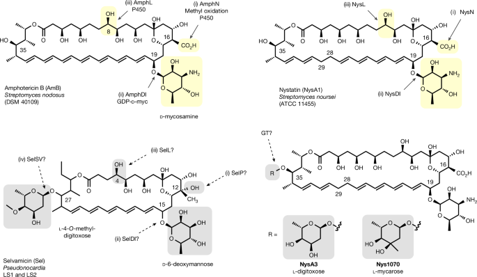
In vitro glycosyltransferase assays for polyene antibiotics
For glycosyltransferase (GT) reactions using TDP-glucose, GT enzyme (5–20 µM), TDP-glucose (2 mM), MgCl2 (1 mM) and polyene substrates—including amphotericin B (AmB), AmB-aglycone, nystatin A1 (NysA1), mycoheptin and dihydromycoheptin—were combined at a final substrate concentration of 100 µM. Reactions were performed in 50 µl of Tris-HCl buffer (50 mM, pH 7.4) and incubated at 30 °C for 24 h.
For TDP sugars with available sugar-1-phosphates, TDP-sugar synthesis was performed before the GT reaction. Deoxythymidine triphosphate (2 mM), sugar-1-phosphate (2 mM), MgCl2 (2.2 mM) and Cps2L (50 µM) were incubated in 50 µl of Tris-HCl buffer (50 mM, pH 7.4) for 24 h at 37 °C in a shaking thermomixer. The temperature was then reduced to 30 °C, and GT (5–20 µM) and polyene substrate (100 µM) were added. Reactions were incubated for an additional 24 h at 30 °C.
For TDP sugars without an available sugar-1-phosphate, the corresponding sugar was used as the starting material. Deoxythymidine triphosphate (2 mM), ATP (2 mM), sugar (2 mM), MgCl2 (2.2 mM), NahK_ATCC15697 or GalkSpe4 (50 µM), and Cps2L (50 µM) were incubated in 50 µl of Tris-HCl buffer (50 mM, pH 7.4) for 24 h at 37 °C in a shaking thermomixer. GT (5–20 µM) and polyene substrate (100 µM) were then added after adjusting the temperature to 30 °C, followed by a further 24-h incubation.
For all one-, two- or three-step assays, reactions were quenched by heating at 95 °C for 5 min and adding one reaction volume of methanol. Proteins were removed by centrifugation, and the supernatants were analysed by analytical reverse-phase high-performance liquid chromatography (RP-HPLC) and liquid chromatography–high-resolution mass spectrometry (LC–HRMS). Analytical HPLC was performed using a Shimadzu ultrahigh-performance liquid chromatography (UHPLC) system equipped with a Kinetex 5 μm XB-C18 100 × 4.6 mm column (Phenomenex). The flow rate was 1 ml min−1, with water and methanol containing 0.1% formic acid and a 60–78% methanol gradient over 12 min. Heptaenes were monitored at 405 nm and pentaenes at 350 nm.
LC–HRMS analyses were performed using an Agilent 1290 Infinity II coupled to a 6560 Ion Mobility quadrupole time-of-flight (Q-TOF) LC–MS system or a 1290 Infinity II HPLC coupled to a 6546 LC–Q-TOF instrument. Separations used a Luna Omega 5 μm 100 × 2.1 mm column (Phenomenex) at 0.5 ml min−1, with water and methanol and a 60–95% methanol gradient over 15 min. Tetraenes, pentaenes and heptaenes were monitored at 304, 350 and 405 nm, respectively.
GT-catalysed reverse glycosylation of polyene antibiotics
Kasufungin B, NysA3 and semipurified selvamicin (25–100 µM) were incubated with TDP (2 mM), MgCl2 (1 mM) and the relevant GT enzyme—KfuSV, NysSV or SelSV—at 25 µM. Reactions were performed in 50 µl of Tris-HCl buffer (50 mM, pH 7.5–8.0) at 30 °C overnight. Reactions were stopped by adding 50 µl of methanol and centrifuged at 10,000 rpm for 5 min. Supernatants were analysed by HPLC, and product masses were confirmed by LC–MS using the conditions described for the in vitro GT glycosylation assays.
MycS3 glycosylation assay using dihydromycoheptin and TDP-l-digitoxose
Reverse glycosylation of NysA3 was used to generate TDP-l-digitoxose. NysA3 (200–300 µM) was incubated with TDP (2 mM), MgCl2 (1 mM) and KfuSV (50 µM) in 50 mM Tris-HCl buffer (pH 7.5–8.0) at 30 °C overnight in a 2-ml reaction. Reactions were quenched with an equal volume of methanol and centrifuged at 10,000 rpm for 5 min. The supernatant was concentrated under vacuum to one-quarter of its original volume, loaded onto silica C18 material (100 Å, 30 μm; 4-ml bed) packed in a small syringe and washed twice with an equal volume of H2O to elute TDP-l-digitoxose. The filtrate was concentrated to one-fifth of its original volume under vacuum and used in MycS3 reactions.
For MycS3 activity assays, dihydromycoheptin (50 µM) was prepared in MycS3 reaction buffer containing MgCl2 (1 mM) and Tris-HCl (50 mM, pH 7.5). The substrate was incubated with 25 µl of concentrated TDP-l-digitoxose filtrate and MycS3 (25 µM) in a 50-µl reaction at 30 °C overnight. Reactions were quenched with 50 µl of methanol, centrifuged at 10,000 rpm for 5 min and analysed by HPLC and LC–MS.
In separate experiments, dried methanolic extracts from S. netropsis (DSM40846) containing crude dihydromycoheptin were resuspended in MycS3 buffer containing MgCl2 (1 mM) and Tris-HCl (50 mM, pH 7.5). The extract was incubated with 25 µl of concentrated TDP-l-digitoxose filtrate and MycS3 (25 µM) at 30 °C overnight in a 50-µl reaction. Reactions were quenched with 50 µl of methanol, centrifuged at 10,000 rpm for 5 min and analysed by LC–HRMS.
HPLC was performed on a Shimadzu Analytical UHPLC fitted with a Kinetex 5 μm XB-C18 100 × 4.6 mm column (Phenomenex), using a 1 ml min−1 flow rate and a 60–78% methanol gradient over 12 min. Both solvents contained 0.1% formic acid, and absorbance was monitored at 350 nm. LC–HRMS used an Agilent 1290 Infinity II coupled to a 6560 Ion Mobility Q-TOF LC–MS or a 6546 LC/Q-TOF system with a Luna Omega 5 μm 100 × 2.1 mm column. The flow rate was 0.5 ml min−1, with a 60–95% methanol gradient over 15 min.
In vitro PcsA enzymatic reaction conditions
PcsA activity was evaluated using 100 µM polyene substrates, including pimaricin, rimocidin, CE-108, NysA1, NysA3, mycoheptin, dihydromycoheptin, kasufungin B and candicidin. Reactions also contained l-glutamine, l-glutamic acid γ-hydroxamate or l-glutamic acid γ-hydrazide (2 mM), ATP (4 mM), MgCl2 (10 mM) and 25 µl of eluted protein in Tris buffer containing 125 mM Tris and 25 mM NaCl (pH 7.0), for a total reaction volume of 50 µl.
l-Glutamine analogues—including l-glutamic acid γ-methylamide, l-glutamic acid γ-ethylamide (l-theanine), l-glutamic acid γ-methyl ester and l-glutamic acid γ-ethyl ester—were also tested; however, no activity was detected. Reactions were incubated overnight at 30 °C, quenched with 50 µl of methanol and centrifuged at 10,000 rpm for 5 min.
HPLC analysis used a Shimadzu Analytical UHPLC equipped with a Kinetex 5 μm XB-C18 100 × 4.6 mm column (Phenomenex), a flow rate of 1 ml min−1 and a 60–78% methanol gradient over 12 min. Water and methanol each contained 0.1% formic acid. LC–HRMS was performed using an Agilent 1290 Infinity II coupled to a 6560 Ion Mobility Q-TOF LC–MS system, with a Luna Omega 5 μm 100 × 2.1 mm column, a flow rate of 0.5 ml min−1 and a 60–80% methanol gradient over 15 min.
Expression of the S. albulus nysSV sugar subcluster in S. albus
The sugar subcluster from S. albulus (DSM40492), designated Sa-nysSV-sub, was amplified from genomic DNA as two fragments. Fragment 1 contained nysSVI, nysSII, nysSV and part of nysSVII; fragment 2 contained the remaining portion of nysSVII, nysSIII and nysSIV. Primer sequences are provided in Supplementary Table 3. The pSET152-ermE* plasmid was linearized with NdeI and EcoRI, and the fragments were assembled by HiFi assembly to generate pSET-ermE*-Sa-nysSV-sub.
The construct was transferred into S. albus J1074, provided by M. Bibb at the John Innes Centre, through E. coli ET12567-mediated conjugation using standard protocols52. This generated the S. albus::Sa-nysSV-sub strain. The empty pSET-ermE* plasmid was transferred in parallel to create the negative-control strain S. albus::pSET-ermE*.
For lysate-based NysA1 assays, both strains were grown in tryptone soy broth (TSB) for 3 days. Seed cultures were transferred at 2% v/v into 10 ml of SG2 medium containing glucose (20 g l−1), yeast extract (5 g l−1) and soytone (10 g l−1; pH 7.2). After 48 h, 1 ml of each culture was centrifuged at 12,000 rpm for 10 min. Pellets were resuspended in lysis buffer at four times the pellet weight. The buffer contained Tris-Cl (50 mM), NaCl (150 mM), glycerol (10%) and MgCl2 (2 mM; pH 7.5). Lysozyme (4–6 mg ml−1) was added, and suspensions were incubated at 30 °C for 30 min, vortexed and centrifuged at 12,000 rpm for 20 min.
A 100-µl aliquot of supernatant was incubated with NysA1 (100 µM) at 30 °C overnight. Reactions were quenched with an equal volume of methanol and centrifuged at 10,000 rpm for 10 min. HPLC was performed using a Shimadzu Analytical UHPLC and Kinetex 5-μm XB-C18 100 × 4.6 mm column at 1 ml min−1, with a 60–78% methanol gradient over 12 min. NysA1 conversion to NysA3 was monitored at 304 nm. LC–HRMS used an Agilent 6546 LC/Q-TOF and 1290 Infinity II HPLC system with a Luna Omega 5 μm 100 × 2.1 mm column, a flow rate of 0.5 ml min−1 and a 60–80% methanol gradient over 15 min.
Expression of kfuSV and Sa-nysSV sugar subclusters in S. nodosus
The sugar subcluster from S. kasugaensis (DSM40819), designated kfuSV-sub, was amplified from genomic DNA as three fragments: fragment 1 contained kfuSVI and kfuSII; fragment 2 contained kfuSIII, kfuSIV and kfuSV; and fragment 3 contained kfuSVII. Primer pairs are listed in Supplementary Table 3. The pSET152-ermE* plasmid was linearized with NdeI and EcoRI, and the fragments were joined by HiFi assembly to create pSET-ermE*-kfuSV-sub.
Similarly, the S. albulus sugar subcluster Sa-nysSV-sub was amplified as two fragments and assembled into pSET-ermE*-Sa-nysSV-sub. Both constructs were separately introduced into S. nodosus DSM40109 using standard conjugation methods52, generating S. nodosus::kfuSV-sub and S. nodosus::Sa-nysSV-sub. The empty pSET-ermE* plasmid was introduced into S. nodosus to produce the negative-control strain S. nodosus::pSET-ermE*.
For polyene production, strains were grown in TSB for 3 days. Seed cultures were transferred at 2% v/v into 50 ml of FSM medium containing fructose (20 g l−1), dextrin (60 g l−1), soya flour (30 g l−1) and CaCO3 (10 g l−1). Cultures were supplemented with Amberlite XAD16N resin (50 g l−1) and incubated with shaking at 30 °C for 7 days. Mycelia and resin were collected by centrifugation, and polyenes were extracted with methanol using one culture volume. A second extraction was performed when additional recovery was required.
S. albulus and S. noursei transcriptome analysis by RT–qPCR
Expression of nysSII, nysSIII and nysSVII was measured using a two-step reverse-transcription quantitative PCR (RT–qPCR) workflow. Starter cultures of S. noursei ATCC 11455 and S. albulus DSM40492 were grown in TSB at 28 °C for 48 h. Starter cultures were transferred at 2% v/v into SPG fermentation medium. After 48 h at 28 °C, 0.5 ml of each culture was harvested, washed with phosphate-buffered saline (PBS) and processed for total RNA using the Monarch Spin RNA Isolation Kit (NEB), including on-column DNase I treatment.
RNA concentration was measured using a NanoDrop instrument (Thermo Scientific), and RNA integrity was assessed with a 4150 TapeStation Analyzer (Agilent). One microgram of purified RNA was used immediately for complementary DNA synthesis with SuperScript IV VILO Master Mix (Thermo Fisher), following the manufacturer’s instructions. cDNA was flash frozen and stored at −80 °C. Primers were designed with the PrimerQuest Tool (IDT) to amplify 80–150-base-pair regions and are listed in Supplementary Table 3.
qPCR was performed with PowerTrack SYBR Green Master Mix (Thermo Fisher) on a Stratagene Mx3000P system. Cycling conditions were 95 °C for 2 min, followed by 40 cycles of 95 °C for 15 s and 60 °C for 60 s, followed by a default dissociation cycle. 16S ribosomal RNA served as the internal control. No-template and no-reverse-transcription controls confirmed the absence of contamination. Three biological replicates were analysed for each gene. Statistical analysis used three unpaired t-tests with an α value of 0.05.
Production and analytical measurement of polyenes from S. albulus and S. noursei
Spores of S. albulus DSM40492 or S. noursei ATCC 11455 were used to inoculate GYM medium. Seed cultures were grown for 3 days at 30 °C and transferred to FSM medium at 2% v/v inoculum in triplicate 50-ml cultures. Amberlite XAD16N resin (50 g l−1) was added to each fermentation, which was incubated with shaking at 30 °C for 7 days.
Mycelia and resin were collected by centrifugation. Polyenes were extracted overnight with one culture volume of methanol at 4 °C in the dark. Extracts were diluted fivefold, and 10 µl was injected for analytical HPLC. Relative NysA1 and NysA3 titres were calculated against an NysA1 standard calibration curve.
Production of polyenes from S. netropsis
Spores of S. netropsis DSM40846 were inoculated into TSB and grown for 2 days at 30 °C. Seed cultures were transferred at 2% v/v into 200 ml of FSM medium in 1-l flasks. FSM contained fructose (20 g l−1), dextrin (60 g l−1), soya flour (30 g l−1) and CaCO3 (10 g l−1). Amberlite XAD16N resin (50 g l−1) was added to improve natural-product recovery. Cultures were incubated with shaking at 30 °C for 7 days, after which mycelia and resin were collected by centrifugation. Polyenes were extracted with methanol at one culture volume and analysed by LC–HRMS.
Production of polyenes from S. kasugaensis
Spores of S. kasugaensis DSM40819 were used to inoculate TSB, and seed cultures were grown for 2 days at 30 °C. Fermentation cultures were prepared by transferring the seed culture at 2% v/v into 200 ml of FSM medium in 1-l flasks. Amberlite XAD16N resin (50 g l−1) was added, and cultures were incubated with shaking at 30 °C for 7 days. Mycelia and resin were collected by centrifugation, and polyenes were extracted twice using methanol at twice the pellet volume.
Polyene purification and NMR structural characterization
Extracts from S. nodosus::kfuSV-sub, S. nodosus::Sa-nysSV-sub, S. albulus, S. netropsis DSM40846 and S. kasugaensis were dried in vacuo. Water (50 ml) was added, and suspensions were incubated at 4 °C for 3 h before centrifugation to collect polyene-containing pellets. Pellets were dissolved in dimethylsulfoxide (DMSO) and purified by semipreparative RP-HPLC using a Shimadzu Prominence HPLC system and a Phenomenex Gemini C18 column (250 × 10 mm; 5 μm).
Before HPLC purification, kasufungin B was partially purified using a Mega BE-C18, 10-g, 60-ml Bond Elut column (Agilent Technologies). The column was washed with up to 70% methanol to remove impurities, followed by 100% methanol to elute pentaene compounds. The eluate was concentrated in vacuo and further purified by semipreparative HPLC.
Mycoheptin, dihydromycoheptin and mandimycin from S. netropsis were purified at 5 ml min−1 using water and methanol with an isocratic 65% methanol phase for 25 min, followed by 95% methanol for 12.5 min. Kasufungin B from S. kasugaensis was purified at 5 ml min−1 using 65% methanol for 11 min, followed by a 66–84% methanol gradient over 12.5 min. AmB-l-digitoxose from S. nodosus::kfuSV-sub was purified using a 70–88% methanol gradient over 30 min. NysA3 from S. albulus was purified using methanol gradients of 40–65% for 5 min and 65–85% for a further 18 min.
Fractions containing the target polyenes were dried in vacuo and dissolved in DMSO-D6 for NysA3 and AmB-l-digitoxose or CD3OD for kasufungin B. One-dimensional (1H and 13C) and two-dimensional NMR experiments—including correlation spectroscopy, heteronuclear single quantum coherence, heteronuclear multiple-bond correlation spectroscopy and rotating-frame nuclear Overhauser effect spectroscopy—were performed at 500 MHz to confirm the structures of mycoheptin, dihydromycoheptin and mandimycin. NysA3, kasufungin B and AmB-l-digitoxose were analysed at 800 MHz.
Antifungal susceptibility testing and IC50 determination
Antifungal susceptibility was measured according to the European Committee on Antimicrobial Susceptibility Testing reference broth microdilution methods, versions 9.3.253 and 7.454. Yeasts—C. albicans ATCC 90028, C. albicans ATCC 200955, C. glabrata NCPF3309, C. auris H.17.157 and Cryptococcus neoformans F10025—were grown on Sabouraud dextrose agar (SDA) at 30 °C for 3 days. Inocula were prepared from five distinct colonies suspended in PBS containing 0.1% Tween and adjusted to 5 × 10⁵ cells ml−1 in sterile distilled water.
Filamentous fungi—including Aspergillus fumigatus A1160, Aspergillus fumigatus cyp51ATR34/L98H, Rhizopus delemar 99-880, Mucor circinelloides 1006Phl, Fusarium oxysporum 9935 and Fusarium solani 9596—were grown in vented SDA T25 flasks at 37 °C for 3 days. Fusarium species were grown at 28 °C for 5 days. Conidia were collected in PBS containing 0.1% Tween, filtered through miracloth and adjusted to 5 × 10⁵ cells ml−1.
Assays were performed in flat-bottom 96-well plates containing 100 µl of 2× Roswell Park Memorial Institute-1640 medium with twofold serial dilutions of antifungal compounds and drug-free controls. Compound stocks were prepared in DMSO. AmB-series compounds were tested at 12.5–0.012 μg ml−1, while pimaricin, NysA1 and NysA3 were tested at 50–0.048 μg ml−1. Each well received 100 µl of the relevant inoculum to give a final concentration of 5 × 10⁴ conidia. Milli-Q water was added to separate wells as a sterility control.
Plates were incubated at 37 °C for 48 h, and optical density at 600 nm (OD600) was measured using a BioTek Synergy 2 SL microplate reader. IC50 values were calculated by fitting variable-slope inhibitory dose–response curves in GraphPad Prism version 10.2.3 (347).
Polyene-induced horse blood haemolysis
Polyene haemolytic activity was measured in horse blood with minor modifications to the method described in reference 28. Polyene dilutions ranging from 0.04 to 350 µM were prepared from 4 mM DMSO stocks. Six microlitres of each dilution was added to 114 µl of PBS containing 2.5% horse blood and incubated at 37 °C for 1 h in 96-well plates. Plates were centrifuged at 4,000 rpm for 15 min, and the absorbance of 100 µl of supernatant was measured at 545 nm.
Compounds were assigned randomly to plate rows. Sixteen final concentrations were tested for AmB, AmB-l-digitoxose, NysA3, Nys31, Nys32, Nys33 and Nys34: 0.04, 0.08, 0.3, 1.25, 10, 20, 30, 40, 50, 60, 70, 80, 90, 100, 150 and 200 µM. Fifteen concentrations were tested for NysA1, Nys11, Nys12, Nys13 and Nys14: 0.6, 1.3, 2.5, 10, 30, 50, 70, 100, 120, 140, 160, 200, 250, 300 and 350 µM. Pim, Pim1, Pim2, Pim3 and Pim4 were tested at 13 concentrations: 10, 30, 50, 70 and 90 µM, together with concentrations from 110 to 350 µM.
Positive and negative controls contained 6 µl of DMSO added to 114 µl of 2.5% horse blood prepared in distilled water or PBS, respectively. Controls were incubated at 37 °C for 1 h and analysed at OD545. Modified polyenes and standards were tested alongside controls in triplicate. EC50 values for 50% haemolysis were calculated using four-parameter logistic curves in GraphPad Prism version 10.2.3 (347).
Human cell-line cytotoxicity testing
A549 human alveolar basal epithelial cells, HEK293 cells and HEPG2 cells were maintained under standard conditions in Gibco DMEM high-glucose, pyruvate medium. Cells were cultured in T75 flasks at 37 °C under 5% CO2. Following trypsinization, A549, HEK293 and HEPG2 cells were seeded into tissue-culture 96-well plates at 10,000, 20,000 and 50,000 cells per well, respectively, and incubated for 24 h in 100 µl per well.
AmB and Nys34 stocks were prepared in DMSO. HEK293 and HEPG2 cells were exposed to final concentrations of 400, 200, 100, 50, 25, 12.5, 6.25 and 3.125 μg ml−1. A549 cells were exposed to 800, 400, 200, 100, 50, 25, 12.5 and 6.25 μg ml−1. Compounds were added to randomly assigned rows, maintaining a final DMSO concentration of 2%.
Cells and medium containing 2% DMSO served as viability controls, while wells containing medium and 2% DMSO without cells served as negative controls. Plates were incubated at 37 °C under 5% CO2 for 18 h. Cell viability was measured using the MTS colorimetric assay with CellTiter 96 AQueous One Solution Reagent (Promega). Absorbance at 490 nm was measured before and 4 h after MTS addition. The pre-treatment absorbance was subtracted from the post-treatment reading. DMSO-treated cells were assigned 100% viability. All experiments were performed in triplicate, and IC50 values were calculated using four-parameter logistic curves in GraphPad Prism.
All cell lines tested negative for mycoplasma and were used as received from Sigma without additional authentication.
Mouse drug tolerability and pharmacokinetic analysis
CD1 mice were purchased from The Jackson Laboratory and transferred to the University of Manchester animal unit under licence PP0175051. Animals were quarantined and acclimatized for at least 1 week before regulated procedures. Mice were housed in individually ventilated cages on a 12-h light–dark cycle at 21 °C ± 2 °C and 40–50% humidity, with nesting material and wood chips. Food and water were provided ad libitum. Mice were checked at the beginning and end of each working day, with an additional check introduced when infection signs or 10% body-weight loss occurred.
Maximum tolerated dose studies used ascending intraperitoneal doses of 1, 2, 5 and 10 mg kg−1, with one mouse receiving each dose. Animals were monitored continuously for 1 h and then hourly for a total of 4 h. Mice were anaesthetized with isoflurane before terminal cardiac puncture and cervical dislocation.
Pharmacokinetic analysis of Nys34 was performed using 20 male and female CD1 mice, comprising 10 animals of each sex and weighing approximately 20 g. Two blood microsamples were collected from each mouse through the tail vein using heparinized capillary tubes. A third terminal cardiac puncture sample was collected to reduce animal numbers in accordance with the 3R principles. Microsamples were collected at 0.5, 1, 2, 4 and 8 h after dosing, with cardiac puncture samples collected at 8 and 24 h according to the scheme in reference 55.
Organ homogenates were extracted with 200% v/w methanol, sonicated at room temperature for 30 min and vortexed vigorously. Extracts were clarified by centrifugation, and extraction was repeated three times. Combined liver extracts were injected directly for LC–MS analysis. Combined lung, spleen and kidney extracts were dried under nitrogen and resuspended in methanol. Lung and spleen samples were resuspended in 50 µl, and kidney samples in 80 µl.
Whole-blood samples were prepared from 5-µl aliquots that were frozen and lyophilized. Dried samples were resuspended in 23 µl methanol, sonicated at room temperature for 30 min and vortexed. Samples were clarified by centrifugation before LC–MS injection. LC–HRMS analysis used a 1290 Infinity II HPLC coupled to an Agilent 6546 LC/Q-TOF system and a Luna Omega 5 μm 100 × 2.1 mm column. The flow rate was 0.5 ml min−1, with water and methanol containing 0.1% formic acid and a 60–95% methanol gradient over 8 min.
Population pharmacokinetic modelling was conducted in R using nlmixr2. Whole-blood concentration–time data were fitted with a one-compartment model incorporating first-order absorption and elimination. Model parameters included the absorption rate constant (ka), clearance (CL) and volume of distribution (V). Exponential random effects described inter-individual variability in CL and V, while residual unexplained variability was represented by a proportional error model. Parameters were estimated using the stochastic approximation expectation–maximization algorithm in nlmixr2. Model performance was assessed using goodness-of-fit plots and residual diagnostics.
To guide dosing-regimen selection, toxicity risk was evaluated using peak concentration (Cmax) and total exposure, expressed as area under the curve. A 10 mg kg−1 dose administered every 8 h for four doses (q8h ×4) was defined as the toxic reference regimen. Monte Carlo simulations based on the population pharmacokinetic model generated 0–32-h peak-concentration distributions. The median simulated Cmax for this regimen was used as the primary toxicity threshold for subsequent screening.
Mouse infection efficacy experiments
For experiments involving Aspergillus fumigatus conidia, 4- to 5-week-old mice were immunocompromised 24 h before infection by subcutaneous administration of Kenalog (triamcinolone acetonide; Bristol Myers Squibb) at 40 mg kg−1. Drinking water was supplemented with neomycin (2 g l−1) at the start of immunosuppression and replaced daily.
Mice were infected intranasally with freshly prepared A. fumigatus CEA10 conidia at 5 × 10⁵ spores in 40 µl sterile saline. Ten CD1 mice—five males and five females—were randomly assigned to each treatment group. Mice received intraperitoneal injections of Nys34 at 5 mg kg−1 or vehicle containing 10% DMSO in saline q8h ×4. Amphotericin B was administered at 1 mg kg−1 every 24 h, beginning 12 h after infection. Body weight and health were monitored at each time point, and mice were euthanized 32 h after the first dose.
To quantify A. fumigatus lung fungal burden, lungs were collected immediately after euthanasia, weighed and transferred to sterile 2-ml microcentrifuge tubes containing 1 ml sterile PBS with 0.1% Tween-20. Lungs were homogenized until no visible tissue fragments remained. Tenfold serial dilutions were prepared in sterile PBS containing 0.1% Tween-20, and 100 µl of each dilution was plated in technical duplicate on SDA containing chloramphenicol (50 µg ml−1). Plates were incubated at 30 °C for 72 h. A. fumigatus colony-forming units (CFUs) were counted, and mean replicate counts were used to calculate total fungal burden. Results were normalized to lung mass and reported as CFU per gram of tissue. Researchers were blinded to sample identity during CFU enumeration.
UV–visible spectroscopy of polyene–sterol binding
Ergosterol stock solutions were prepared at 100 mM in chloroform and diluted in DMSO to 1 mM. AmB, NysA1 and Nys34 were prepared as 1 mM DMSO stocks. Polyene–sterol complexes were prepared in triplicate in 96-well plates by combining the stocks at defined molar ratios from 1:0 to 1:5, as detailed in Supplementary Table 6. Complexes were incubated at room temperature for 30 min before recording UV–visible absorption spectra with a microplate spectrophotometer. NysA1 derivatives were scanned from 280 to 340 nm, and AmB derivatives from 380 to 440 nm, using a 1-nm wavelength interval6.
Effect of sterol precomplexation on polyene antifungal activity
Ergosterol was recrystallized from ethanol and prepared as a stock solution in CHCl3. Appropriate quantities were dispensed, the solvent was removed under nitrogen and samples were dried completely under vacuum overnight. Polyene DMSO solutions were added to solid ergosterol at a 5:1 ergosterol-to-polyene molar ratio. Suspensions were gently vortexed, heated at 80 °C for 1 h and cooled to room temperature to allow complex formation.
S. cerevisiae was grown on SDA for 3 days at 30 °C. Five distinct colonies larger than 1 mm were suspended in PBS containing 0.1% Tween-20 and adjusted to an OD600 of 0.5. The suspension was then diluted tenfold to obtain 5 × 10⁵ cells ml−1. For A. fumigatus A1160, spores were grown on SDA at 37 °C, harvested in PBS containing 0.1% Tween-20 and adjusted to 5 × 10⁵ spores ml−1 in sterile distilled water.
AmB, NysA1 and Nys34 were prepared as 2.7 mM, 5.4 mM and 5.4 mM DMSO stock solutions, respectively, with or without ergosterol precomplexation. Stocks were serially diluted twofold in DMSO and then diluted 100-fold in 2× YPD medium for S. cerevisiae or RPMI medium for A. fumigatus. One hundred microlitres of each medium–polyene solution was added to 96-well plates containing 100 µl of fungal inoculum in triplicate. Plates were incubated at 30 °C for 24 h before OD600 measurement. Control wells containing 0.5% DMSO confirmed viability, and water-only inoculum wells served as sterility controls54.
Reporting summary
Additional information about the research design, experimental procedures and reporting standards is available in the Nature Portfolio Reporting Summary linked to this article.
Source: www.nature.com