


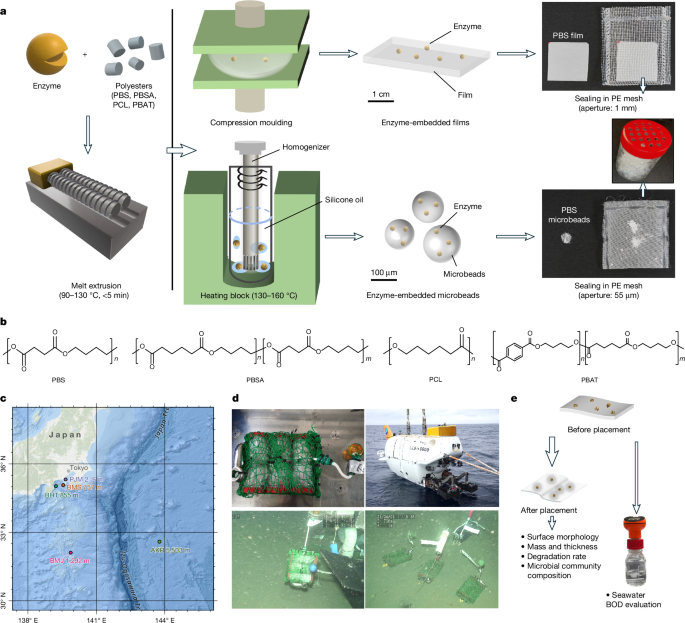
The study examined enzyme-embedded polyester films and microbeads under laboratory, marine and deep-sea conditions. Researchers prepared films from PBS, PBSA, PCL, PBAT and P3HB, incorporating HiC or CaLB enzymes, and evaluated their physical properties, biodegradation, microbial communities, surface morphology, enzymatic degradation products and performance under pressures of up to 25 MPa. The work included deep-sea deployment using the HOV Shinkai 6500 and Kaimei-ROV, biochemical oxygen demand testing, sequencing of 16S rRNA genes and a range of mechanical and chemical analyses.
Sample preparation
PBS, PBSA, PCL, PBAT, P3HB and polyethylene terephthalate (PET) pellets were purchased from or supplied by commercial sources listed in Supplementary Table 1a. HiC (Novozyme 51032, 15,000 LU g−1) and CaLB (Lipozyme CALB L, 5,000 LU g−1) were purchased from Wako Fujifilm. Enzyme powders were prepared as previously described16,18.
To produce enzyme-free films, PBS, PBSA, PCL, PBAT and P3HB pellets were dried at 40–50 °C under reduced pressure for at least 12 h to remove residual moisture. Approximately 2–3 g of each dried polymer was fed into a counter-rotating twin-screw micro-extruder (Thermo Scientific HAAKE MiniLab) running at 120 rpm. The processing temperatures were 90 °C for PCL, 100 °C for PBSA, 130 °C for PBS and PBAT, and 180 °C for P3HB. Residence time in the extruder was limited to 3 min to reduce thermal degradation and further enzyme denaturation.
The molten polymer was extruded through a 1 × 2-mm rectangular die and cooled to room temperature. Approximately 300 mg of extrudate was then compression-moulded for 30 s at 2 MPa and the relevant extrusion temperature, followed by quenching in ice water for 10 s. The combined extrusion and pressing time was no more than 3.5 min. Films were approximately 100–300 µm thick and were stored in a desiccator at room temperature for at least 48 h and up to one month.
Enzyme-embedded films were made by adding HiC or CaLB to the polymer pellets at a loading of 0.1 wt%; loadings of up to 3 wt% were used for samples intended for deep-sea deployment. Dried pellets were thoroughly mixed with enzyme powder and melt-extruded using the same procedure and temperature settings as the enzyme-free polymers. After 3 min of continuous mixing, the extrudate was cooled. Approximately 300 mg of the enzyme-polymer blend was compression-moulded for 30 s at 2 MPa, then quenched in ice water and stored using the same protocol.
To prepare enzyme-embedded PBS, PBSA and PBAT microbeads, 4 ml of silicone oil was placed in a stainless-steel tube and heated to the designated melt-processing temperature in an aluminium-block thermostatic bath (DTU-1C, TAITEC) (Extended Data Fig. 1). Pre-cut pieces of enzyme-embedded polymer extrudate weighing 0.2–0.3 g were added to the oil and melted for 3 min. A Microtec homogenizer fitted with an NS-7 generator shaft (7.0-mm diameter × 100 mm) dispersed the molten polymer at 12,000–24,000 rpm for 60 s. The tube was then rapidly cooled in a 4 °C ice-water bath.
The resulting microbeads, suspended in silicone oil, were separated by three cycles of washing with hexane and centrifugation. The beads were recovered by vacuum filtration through a 0.45-µm PTFE membrane, resuspended in hexane and filtered through a 250-µm polyethylene mesh. They were washed twice with hexane, air-dried for one day and stored at 4 °C. PCL microbeads could not be produced using the homogenization-in-silicone-oil method.
Sample thickness was measured at five locations before and after environmental degradation tests using a digital micrometer (Mitsutoyo), and the measurements were averaged. For melt-pressed films, the five locations were the four corners and centre of a 4 × 4-cm film. The mean and standard deviation were calculated. Dimensions were measured to two decimal places in centimetres using a digital calliper (Shinwa), and mass was measured with an electronic balance.
Films and microbeads were sealed in polyethylene mesh with apertures of 1 mm and 55 μm, respectively, and placed in PET chambers with through holes. The 55-μm aperture was selected to retain particles while minimizing diffusion limitations and enclosure artefacts during long-term marine exposure. Because the microbeads were smaller than the mesh openings, some particle loss could occur, preventing reliable gravimetric determination of mass loss.
Enzyme-free and enzyme-embedded polyester samples were stored in separate containers to prevent cross-contamination. Films containing CaLB and HiC were also placed in separate containers when deployed simultaneously. Each sample type had at least five total replicates, with a minimum of four used for the main measurements and one reserved for additional analyses.
Sample deployment and recovery
Figure 1c was created from geospatial data obtained from multiple sources using ArcGIS Pro. The base map was provided by Esri, with additional geographic information from TomTom, Garmin, FAO, NOAA, USGS, GEBCO and NaturalVue.
Deep-sea samples were deployed and recovered using either the HOV Shinkai 6500 or remotely operated vehicles (ROVs), including Kaimei-ROV. To prevent contamination from upper-layer seawater during transit, sample chambers were placed in a box covered with a lid.
After recovery, the sample chambers were dismantled promptly. One specimen from each sample set was immersed in 4% formaldehyde prepared in 0.1 M phosphate buffer (pH 7.4) containing 150 mM NaCl for scanning electron microscopy (SEM). Another specimen was frozen at −80 °C for physicochemical analyses. Recovered samples were defrosted in the laboratory, sonicated in distilled water for 30 s to remove surface biofilm and dried under vacuum overnight at room temperature.
Mass was measured in grams to four decimal places using an electronic balance. Mass and dimensions were measured, and values reported for films represent averages from four or five samples. Data are presented as mean ± s.d. Mass loss was calculated as follows, where M represents mass:
Mass loss (wt%) = ((Minitial − Mfinal) / Minitial) × 100
At BMS, deep-sea water next to the sample chambers was collected from the sample box of Shinkai 6500. Immediately after retrieval, the water was stored at 4 °C.
16S rRNA gene amplicon sequencing
16S rRNA gene amplicon sequencing and subsequent analyses were conducted in collaboration with Bioengineering Lab. Co., Ltd. Genomic DNA was extracted from polyester samples by adding Lysis Solution F (Nippon Gene), followed by mechanical disruption with a Shake Master Neo (bms) at 1,500 rpm for 2 min. The lysate was incubated at 65 °C for 10 min and centrifuged at 12,000g for 2 min. The supernatant was purified using the Lab-Aid 824s DNA Extraction Kit (ZEESAN).
DNA concentration was measured with the Synergy LX (Agilent Technologies) and QuantiFluor dsDNA System (Promega). Libraries were prepared using a two-step tailed PCR protocol. The first PCR reaction had a total volume of 10 µl and contained 5.0 µl of 2× PCR Buffer for KOD FX Neo, 2.0 µl of 2 mM dNTPs, 0.2 µl each of 10 µM forward primer 515 F (5′-ACACTCTTTCCCTACACGACGCTCTTCCGATCTNNNNNGTGCCAGCMGCCGCGGTAA-3′) and reverse primer 806 R (5′-GTGACTGGAGTTCAGACGTGTGCTCTTCCGATCTGGACTACNVGGGTWTCTAAT-3′), 1.0 µl of template DNA, 0.2 µl of KOD FX Neo (TOYOBO) and 1.4 µl of nuclease-free water.
The first PCR cycling conditions were 94 °C for 2 min, followed by repeated cycles of 98 °C for 10 s, 50 °C for 30 s and 68 °C for 30 s, with a final extension at 68 °C for 7 min. PCR products were purified using VAHTS DNA Clean Beads (Vazyme) at 1.0× the reaction volume.
The second 10-µl PCR reaction used the same reagents, but included 0.5 µl each of 5 µM forward primer 2ndF (5′-AATGATACGGCGACCACCGAGATCTACAC-Index2-ACACTCTTTCCCTACACGACGC-3′) and reverse primer 2ndR (5′-CAAGCAGAAGACGGCATACGAGAT-Index1-GTGACTGGAGTTCAGACGTGTG-3′). Cycling conditions were 94 °C for 2 min, repeated cycles of 98 °C for 10 s, 50 °C for 30 s and 68 °C for 30 s, and a final extension at 68 °C for 2 min. The final libraries were purified again with VAHTS DNA Clean Beads.
Libraries were quantified using the Synergy H1 and QuantiFluor dsDNA System. Quality was checked with the Fragment Analyzer and dsDNA 915 Reagent Kit (Agilent Technologies). Sequencing was performed on a NextSeq 1000 system using the P1 reagents (600 cycles) kit (Illumina).
Primer-matching reads were extracted with the FASTX-Toolkit (v0.0.14) fastx_barcode_splitter tool, trimmed with FASTX-Toolkit fastx_trimmer and quality-filtered with sickle (v1.33). Reads with Q < 20 or lengths below 130 bp were discarded. Paired-end reads were merged using FLASH (v1.2.11) with a junction sequence length of 250 bp, read length of 230 bp and overlap length of 10 bp.
QIIME2 (v2024.10) with the DADA2 plugin was used to remove chimeric and noisy sequences and generate amplicon sequence variants (ASVs). Phylogeny was generated using the feature-classifier plugin and the EzBioCloud 16S database51, updated on 10 July 2017 (https://help.ezbiocloud.net/ezbiocloud-16s-database/). Results were visualized using OriginPro (v2022).
Because samples were prioritized for mass-loss measurements, 16S rRNA sequencing was performed on one sample for each condition, without independent biological replication.
BOD tests of HiC-embedded polyester films
Biochemical oxygen demand (BOD) analyses used seawater and sediment collected from the port of JAMSTEC (PJM) (35° 19.18′ N, 139° 39.07′ E) in March 2025. Immediately after collection, seawater was transferred to plastic tanks and stored at ambient temperature with loosely closed lids for one week. The supernatant was then filtered through qualitative filter paper (Advantec No. 1) and kept at room temperature overnight before use.
All BOD measurements were conducted under aerobic conditions in a seawater-based medium using an OxiTop-iDS system (WTW), following ASTM D6691-17 guidelines. Experiments ran at 25 °C with continuous stirring for one month. Film samples measuring 0.5 × 0.5 cm, with a theoretical oxygen demand (THOD) of 20 mg O2 per bottle, were placed in 250-ml BOD bottles containing 100 ml of processed seawater.
Each bottle received a mineral solution with final concentrations of 0.109 g l−1 KH2PO4, 21.75 mg l−1 K2HPO4, 33.4 mg l−1 Na2HPO4•2H2O and 0.502 g l−1 NH4Cl, along with 5 mg l−1 allylthiourea.
The tests used 0.1 wt% HiC-embedded PBS, PBSA, PCL and PBAT films. Polyester films without embedded enzyme and cellulose filter paper (Whatman No. 42) served as controls. Blank measurements containing seawater only were also performed. Each test condition had three replicates. Biodegradation was calculated as:
Biodegradability (%) = ((BODt − BODb) / THOD) × 100
BODt is the BOD of the test solution in mg l−1; BODb is the BOD of the blank in mg l−1; THOD is the theoretical oxygen demand of the sample in mg l−1; and biodegradability is reported as a percentage.
SEM observation
Recovered plastic specimens were fixed in 4% formaldehyde prepared in 0.1 M phosphate buffer (pH 7.4) containing 150 mM NaCl, then rinsed in deionized water. Samples were dehydrated through an ethanol series of 30%, 50%, 70%, 90% and 100%, with each step lasting 10 min at room temperature. They were then subjected to two substitutions with tert-butyl alcohol and freeze-dried.
Two sputter-coating and imaging protocols were used. In the first, samples were coated with gold using an MSP-1S magnetron sputter coater (Vacuum Device) and observed in secondary-electron mode with a JCM-7000 microscope (JEOL) at an accelerating voltage of 5 or 10 kV. In the second, samples were coated with platinum for 30 s using an E-1030 ion-sputtering device (Hitachi), then imaged with an S-4800 field-emission scanning electron microscope (Hitachi) at an accelerating voltage of 1.0 kV.
Laboratory pressure-vessel simulation experiments
Deep-sea water collected at BMS was autoclaved at 120 °C for 20 min. Square PBSA and PCL films, each measuring 2 cm × 2 cm, were placed in individual zip-top polyethylene bags. Each bag received 10 ml of a CaLB solution at 100 LU ml−1, prepared with the autoclaved deep-sea water. Air was expelled before the bags were tightly sealed and transferred to a pressure vessel filled with deionized water.
Two pressure vessels were used. One was pressurized to 10 MPa and the other to a maximum of 25 MPa. Both were maintained at 4 °C for one month. The films were then removed, air-dried and weighed to assess mass changes. A non-pressurized control experiment was conducted in parallel at 4 °C. Each dataset contained two replicate samples.
Tensile testing and compression testing of individual microbeads
Film tensile properties were evaluated at room temperature with a universal testing machine (EZ-LX, Shimadzu). The strain rate was 200 mm min−1, and the initial gauge length was 10 mm. Elongation at break, Young’s modulus and maximum tensile strength were calculated from the average of five dumbbell-shaped specimens prepared according to Japanese Industrial Standard (JIS) K 6251-5 1/3. Specimens were prepared two days before testing and stored in a desiccator under ambient conditions.
Microbeads were compression-tested with a flat-punch indenter on a micro compression tester (MCT-211; Shimadzu). Bead sizes were determined using an optical microscope attached to the tester. A compressive force from 0 to 200 mN was applied to each bead at an increasing rate of 4.8 mN s−1.
Under JIS Z8844:2019, which covers fracture-strength and deformation-strength measurements for microbeads, compression strength was assessed from the stress at 10% strain. Because plastic microbeads are soft and do not have a definite crush point, the study did not use a distinct crushing force. Nine individual microbeads smaller than 100 μm were selected and investigated.
HPLC analysis of enzymatic degradation intermediates
PBAT films containing 0.5 wt% HiC were placed in a glass container with 2 ml of 100 mM phosphate buffer at pH 7.5. Samples were incubated at 45 °C for 96 h. After incubation, the reaction solution was collected and heated at 100 °C for 15 min to denature the enzyme. Enzymatic degradation intermediates were analysed using a high-pressure-gradient HPLC system.
The system comprised a CBM-20A communication bus module, a DGC-20A3R degasser for the water-phase pump unit, an LC-20AD liquid chromatograph, a SIL-20A autosampler, an SPD-20A UV/Vis detector and a CTO-20A column oven, all from Shimadzu. UV detection was performed at 254 nm.
Separation used a reversed-phase Asahipak ODP-50 4E column and an Asahipak ODP-50G 4A guard column, both from Shodex. The mobile phase consisted of water containing 1% formic acid and acetonitrile. The elution gradient changed from a 15:85 ratio to 85:15 over 40 min. The flow rate was 1 ml min−1, and the column temperature was 40 °C.
Hydrolysis products were identified and quantified using synthesized mono(4-hydroxybutyl) terephthalate (MHBT) and bis(4-hydroxybutyl) terephthalate (BHBT) as reference standards, together with terephthalic acid. All compounds were analysed using the same HPLC system. Adipic acid was quantified with an ion-exclusion chromatography column (SH1011, Shodex), using 0.01 N sulfuric acid as the mobile phase at a flow rate of 0.6 ml min−1 and a temperature of 60 °C.
Data analysis and visualization
Data organization, statistical analysis and visualization were performed using Microsoft Excel for Microsoft 365 MSO (v2509, build 16.0.19231.20138, 64-bit), OriginPro 2022 SR1 (v9.9.0.225, 64-bit, Academic) and Microsoft PowerPoint for Microsoft 365 MSO (v2509, build 16.0.19231.20138, 64-bit). Maps were prepared with ArcGIS Pro 2024. Three-dimensional illustrations were created using Shapr3D (iPad educational version, 2024).
Reproducibility and statistical analysis
No statistical methods were used to predetermine sample sizes. The study did not use blinding or randomization. Statistical analyses were performed for quantitative datasets as indicated in the corresponding figures. Two-tailed Student’s t-tests were used for pairwise comparisons, as indicated in the figures and Supplementary Tables.
For deep-sea deployment experiments, each sample type was prepared with five or six independent specimens whenever possible. Four or five independent specimens were used for quantitative measurements, and one was reserved for additional analyses. Mass and degradation-rate data for films are presented as mean ± s.d. from independent specimens, as indicated.
Statistical significance was denoted as NS (not significant; P ≥ 0.05), *P < 0.05, **P < 0.01 and ***P < 0.001. Exact sample sizes (n), test statistics, degrees of freedom and exact P values are provided in Supplementary Tables 11–18.
For macroscopic photographic documentation, four or five independent specimens were prepared and visually inspected for each condition. Similar appearances were observed across specimens, and representative photographs are shown in the figures. SEM images are also representative. Because sample availability was limited, one specimen was examined for each condition. For films, at least two distinct areas of the same specimen were observed, with similar surface features found across areas. For microbeads, three independent particles were examined, and similar morphological features were observed among them.
For 16S rRNA gene amplicon sequencing, one sample was analysed for each condition because sample availability was prioritized for mass-loss measurements; there was no independent biological replication. BOD measurements used three independent experimental units, consisting of individual bottles, for each condition. Laboratory pressure-vessel simulations included two independent film specimens per condition. Tensile testing used five independent dumbbell-shaped film specimens per condition. Microbead compression testing used five independent microbeads per condition.
Reporting summary
Further information on the research design is available in the Nature Portfolio Reporting Summary linked to the article.
Source: www.nature.com