


Human brain development begins before birth through a complex process of cell selection and specialization. At the center of this process are radial glia, a unique type of neural stem cell that helps build many of the structures that define the human brain.
Radial glia produce large numbers of neurons and help generate the cells that form the cerebral cortex, the brain region responsible for thinking, memory, language, and other advanced functions. These neural stem cells may also contribute to the remarkable expansion of the human cortex compared with that of other species. Although most radial glia disappear before birth, similar cells can reappear in brain tumors for reasons scientists are still working to understand.
“Radial glia are the coolest cells that have ever existed,” said Aparna Bhaduri, assistant professor of biochemistry at UCLA’s David Geffen School of Medicine. “They are really key to what makes us human. But they are also involved in many neurodevelopmental and neuropsychiatric disorders, not just cancer. Understanding how they make decisions is one way to begin understanding how these conditions develop.”
Two new studies published in Cell and Science reveal how radial glia make developmental decisions. Bhaduri and her colleagues found that these neural stem cells respond to two very different types of information: metabolic signals that show how nutrients are being processed and physical signals delivered by other cells in the developing brain. Together, the findings offer new insight into how the human cerebral cortex produces its diverse range of cell types.
Metabolism helps guide neural stem cell development
In the Cell study, researchers created a detailed map of metabolism in the developing human cerebral cortex. The project brought together Bhaduri’s laboratory and the laboratory of Heather Christofk. It was led by co-first authors Jessenya Mil and Jose Soto.
To build the metabolic atlas, the team analyzed donated human brain tissue as well as brain organoids grown from stem cells. The findings revealed that metabolism does more than provide background support for brain development. It can actively influence which types of cells neural stem cells produce.
Researchers found that radial glia rely heavily on the pentose phosphate pathway, a metabolic process that uses glucose to produce molecules needed by rapidly dividing cells.
When scientists reduced glucose availability or disrupted the pentose phosphate pathway, radial glia changed their developmental behavior. The cells began producing more inhibitory neurons and other cell types that typically emerge later in brain development.
“What surprised me was that metabolism is not just a passive process happening in the background,” said Bhaduri, a member of the UCLA Broad Stem Cell Research Center and the UCLA Health Jonsson Comprehensive Cancer Center. “We can actually influence how neural stem cells make developmental decisions.”
The findings could help scientists investigate how maternal nutrition, metabolic disorders, and other environmental factors affect fetal brain development. The metabolic atlas also provides a valuable resource for researchers studying how metabolism shapes the developing human brain.
Thalamic signals reach radial glia early in development
The second study, published in Science and led by first author Claudia Nguyen, examined a completely different source of developmental information. Researchers focused on signals from the thalamus, a structure deep within the brain that helps relay information throughout the nervous system.
Scientists have known for years that thalamic neurons send long projections toward the cerebral cortex. These wire-like extensions eventually form connections with specific cortical neurons. However, anatomical studies have shown that in humans, thalamic projections reach the cortex long before their final neural connections are established.
This raised an important question: Why do these fibers arrive so early?
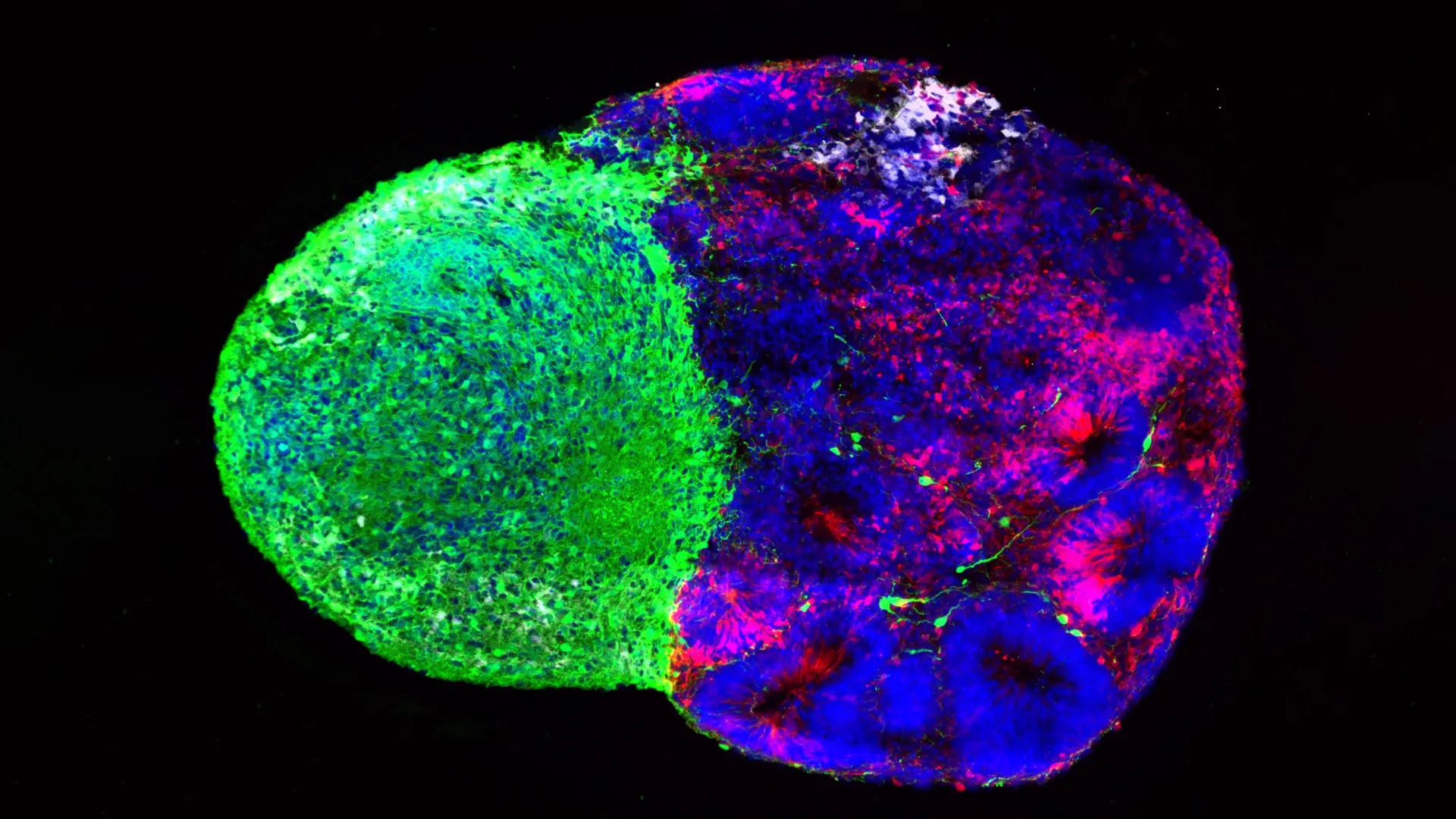
UCLA researchers found part of the answer by using human stem cell-derived brain “assembloids.” During early brain development, thalamic projections make direct physical contact with radial glia.
This contact changed the behavior of the neural stem cells and increased the production of excitatory neurons, which are the primary signal-carrying neurons in the cerebral cortex. The effect was especially strong in upper-layer neurons, a population that is particularly expanded in the human brain.
“We already knew that these projections influence cortical development,” Bhaduri said. “What we specifically discovered is that this effect occurs through a physical connection between the projections and radial glia. This previously unidentified point of contact is likely absent in rodents.”
Autism-related gene influences cortical development
The researchers connected this physical interaction to NRXN1, a gene involved in forming connections between neurons. Mutations in NRXN1 have previously been associated with autism spectrum disorder.
To study the gene’s role, the research team created brain cell aggregates using cells from patients with NRXN1 mutations. In these models, altered thalamic signals behaved differently from signals generated by unaffected cells.
These changes affected the number of neural stem cells and altered the balance of neurons they produced. The results provide scientists with a potential model for studying how early neurodevelopmental disorders disrupt formation of the cerebral cortex.
The developing brain is constantly communicating
Although the two studies examined different mechanisms, they point to the same broader conclusion: radial glia do not make developmental decisions in isolation. Their behavior is continuously shaped by signals from the surrounding environment, including metabolic activity and physical interactions with other brain cells.
The studies also highlight how brain organoids and assembloids are transforming research into human brain development. Just over a decade ago, scientists had few practical tools for directly investigating the unique behavior of human neural stem cells.
Today, brain organoids and related laboratory models can reproduce important features of human brain development. They also allow researchers to investigate conditions that cannot be fully studied using animal models alone.
Bhaduri hopes the findings will encourage researchers to view metabolism and physical cell connections as active drivers of brain development rather than passive background processes.
“Ultimately, these studies provide an inside view of how radial glia make decisions,” she said. “Understanding these decisions is the first step toward learning how similar stem cell programs operate during normal brain development, disease vulnerability, and potentially the formation of brain tumors.”
This research was supported by the National Institutes of Health, the National Science Foundation, the Brain and Behavior Research Foundation, the Alfred P. Sloan Foundation, the Rose Hills Foundation, the Esther A. Joseph Klingenstein Foundation, the Simons Foundation, the Chan Zuckerberg Initiative, the NIH BRAIN Initiative Cell Atlas Network, the International Foundation for Ethics Research, the UCLA Broad Stem Cell Research Center Stem Cell Research Training Program, and the UCLA Health Jonsson Comprehensive Cancer Center and UCLA Broad Stem Cell Research Center Abron Scholars Program.
Source: www.sciencedaily.com