


New Flexible Brain Implant Records Neural Activity and Delivers Drugs at Multiple Depths
A new type of brain implant could give scientists a more precise way to study the brain and may eventually contribute to treatments for neurological conditions such as epilepsy.
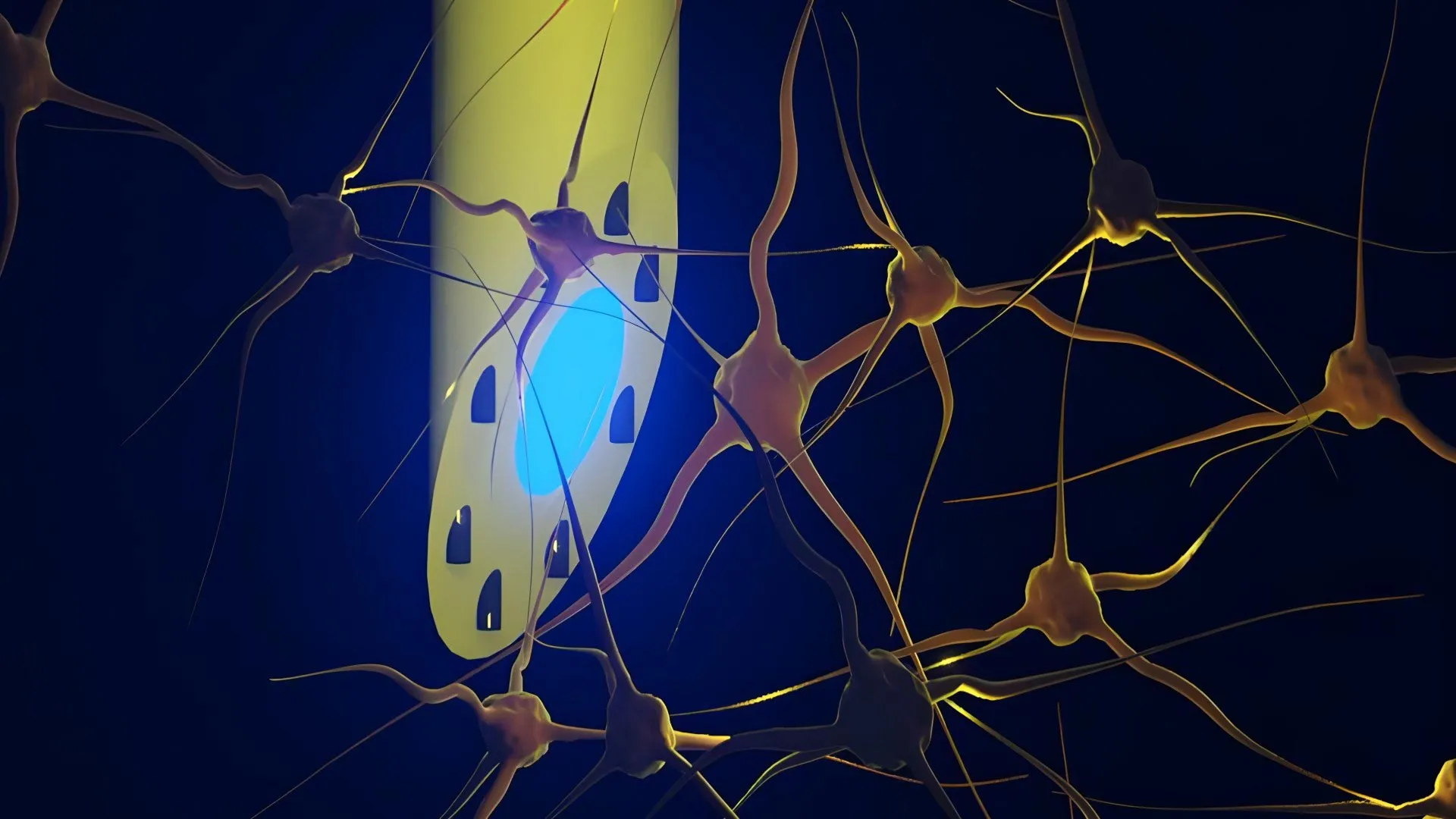
Researchers at DTU, the University of Copenhagen, University College London and other institutions have developed long, needle-thin brain electrodes containing microscopic channels. The device, known as a microfluidic Axialtrode, or mAxialtrode, is designed to provide multiple functional points along the length of a single implant.
This design allows researchers to record neural activity and deliver drugs to specific locations across different regions and layers of the brain. The research results were published in the magazine Cutting Edge Science.
A multifunctional tool for brain research
For now, the mAxialtrode is primarily intended as a research tool. Scientists could use it to investigate how signals travel through different layers of the brain during processes involved in epilepsy, memory and decision-making.
In the long term, the researchers say the technology may also have therapeutic applications. One possibility would be to use the device to apply electrical or optical stimulation to selected areas of the brain while simultaneously delivering drugs to precise locations.
Kunyan Sui, a postdoctoral researcher who developed the mAxialtrode concept with Associate Professor Christos Marcos, says one of its main advantages is the ability to combine multiple functions within a single implant. This could allow researchers to perform more precise experiments while reducing the need for multiple devices inserted into the brain.
“Most current brain implants are based on hard materials such as silicone, which can irritate the brain and cause an inflammatory response within the tissue. The new implant differs in that it is made of soft, plastic-like optical fibers and has a specially angled tip. This makes the implant smaller and reduces the damage caused when it is placed in the brain,” Kunyan Sui says.
Sui cautioned that the technology is still far from routine clinical use. Extensive trials, further development and regulatory approval will be required before it can be used to treat patients.
Why traditional brain electrodes have limitations
Brain researchers currently use flat-ended optical fibers in many experiments. These thin fibers, made of glass or plastic, can carry light deep into the brain and are frequently used in optogenetics, a technique that uses light to activate specific nerve cells.
However, traditional optical fibers have important limitations. They typically interact with brain tissue only at their ends, allowing researchers to stimulate or monitor one location at a time.
The outermost end is called the distal end, or “nose,” of the fiber. Light is emitted and contact with brain tissue occurs only at this point. As a result, scientists may be limited to measuring or stimulating one brain layer at a time, even though many brain functions rely on communication between multiple layers and deeper structures.
How the mAxialtrode brain implant works
The needle-thin mAxialtrode begins as a much larger polymer rod. Researchers heat the material and draw it into very thin fibers. The process is similar to making extremely thin sugar threads, but with much greater precision.
A photoconductive core runs through the center of the fiber. Around it are eight microscopic channels that can transport fluids. The channels can also hold very thin metal wires used to measure electrical activity in the brain.
The finished fiber has a diameter of less than 0.5 millimeters. It is also highly flexible, allowing it to move along with brain tissue rather than pushing against it. This difference in stiffness may be important because stiff implants can cause an inflammatory response if they remain in the brain for long periods.
Brain implant tested in living mice
The researchers tested the system both in the laboratory and in vivo, meaning in living mice. Electrodes were implanted in the animals’ brains and connected to a light source, a recording device and a small pump used to deliver fluid.
The experiments showed that the device could use both blue and red light to stimulate nerve cells. At the same time, researchers were able to record electrical activity from shallow and deep regions of the brain, including the cerebral cortex and hippocampus.
The team also injected different substances at different depths, with injection points spaced approximately 3 millimeters apart. All of these measurements and forms of stimulation were performed using a single lightweight fiber, and the mice were able to carry it without exhibiting obvious discomfort.
Potential applications in epilepsy and neuroscience
The in vivo experiments and neurophysiological validation were carried out in close collaboration with Associate Professor Rune W. Berg from the University of Copenhagen and Associate Professor Rob C. Wykes from University College London. Their contributions included expertise in analyzing neural circuits and models related to epilepsy.
The research team is currently working to patent the technology behind the brain electrodes. Scientists are also investigating what would be required to begin testing the device on patients in a clinical setting.
Source: www.sciencedaily.com